Crystal structure of the Mor protein of bacteriophage Mu, a member of the Mor/C family of transcription activators.
Kumaraswami, M., Howe, M.M., Park, H.W.(2004) J Biological Chem 279: 16581-16590
- PubMed: 14729670 Search on PubMed
- DOI: https://doi.org/10.1074/jbc.M313555200
- Primary Citation Related Structures:
1RR7 - PubMed Abstract:
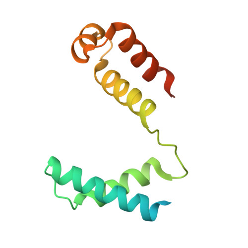
Transcription from the middle promoter, Pm, of bacteriophage Mu requires the phage-encoded activator protein Mor and bacterial RNA polymerase. Mor is a sequence-specific DNA-binding protein that mediates transcription activation through its interactions with the C-terminal domains of the alpha and sigma subunits of bacterial RNA polymerase. Here we present the first structure for a member of the Mor/C family of transcription activators, the crystal structure of Mor to 2.2-A resolution. Each monomer of the Mor dimer is composed of two domains, the N-terminal dimerization domain and C-terminal DNA-binding domain, which are connected by a linker containing a beta strand. The N-terminal dimerization domain has an unusual mode of dimerization; helices alpha1 and alpha2 of both monomers are intertwined to form a four-helix bundle, generating a hydrophobic core that is further stabilized by antiparallel interactions between the two beta strands. Mutational analysis of key leucine residues in helix alpha1 demonstrated a role for this hydrophobic core in protein solubility and function. The C-terminal domain has a classical helix-turn-helix DNA-binding motif that is located at opposite ends of the elongated dimer. Since the distance between the two helix-turn-helix motifs is too great to allow binding to two adjacent major grooves of the 16-bp Mor-binding site, we propose that conformational changes in the protein and DNA will be required for Mor to interact with the DNA. The highly conserved glycines flanking the beta strand may act as pivot points, facilitating the conformational changes of Mor, and the DNA may be bent.
- Department of Molecular Sciences, University of Tennessee Health Science Center, Memphis, Tennessee 38163, USA.
Organizational Affiliation: