1ROA | pdb_00001roa

Structure of human cystatin D


Experimental Data Snapshot

  • Method: X-RAY DIFFRACTION
  • Resolution: 1.80 Å
  • R-Value Free: 
    0.278 (Depositor), 0.272 (DCC) 
  • R-Value Work: 
    0.250 (Depositor), 0.247 (DCC) 
  • R-Value Observed: 
    0.250 (Depositor) 

Starting Model: experimental
View more details

wwPDB Validation 3D Report Full Report

Validation slider image for 1ROA

This is version 1.6 of the entry. See complete history

Literature

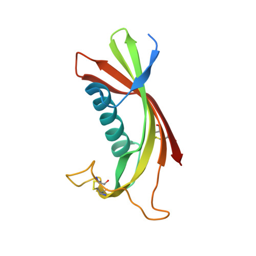
Crystal structure of human cystatin D, a cysteine peptidase inhibitor with restricted inhibition profile.

Alvarez-Fernandez, M.Liang, Y.H.Abrahamson, M.Su, X.D.

(2005) J Biological Chem 280: 18221-18228

  • DOI: https://doi.org/10.1074/jbc.M411914200
  • Primary Citation Related Structures: 
    1RN7, 1ROA

  • PubMed Abstract: 

    Cystatins are natural inhibitors of papain-like (family C1) and legumain-related (family C13) cysteine peptidases. Cystatin D is a type 2 cystatin, a secreted inhibitor found in human saliva and tear fluid. Compared with its homologues, cystatin D presents an unusual inhibition profile with a preferential inhibition cathepsin S > cathepsin H > cathepsin L and no inhibition of cathepsin B or pig legumain. To elucidate the structural reasons for this specificity, we have crystallized recombinant human Arg(26)-cystatin D and solved its structures at room temperature and at cryo conditions to 2.5- and 1.8-A resolution, respectively. Human cystatin D presents the typical cystatin fold, with a five-stranded anti-parallel beta-sheet wrapped around a five-turn alpha-helix. The structures reveal differences in the peptidase-interacting regions when compared with other cystatins, providing plausible explanations for the restricted inhibitory specificity of cystatin D for some papain-like peptidases and its lack of reactivity toward legumain-related enzymes.


  • Organizational Affiliation
    • Department of Clinical Chemistry, Institute of Laboratory Medicine, Lund University, SE-221 85 Lund, Sweden.

Macromolecule Content 

  • Total Structure Weight: 13.93 kDa 
  • Atom Count: 993 
  • Modeled Residue Count: 111 
  • Deposited Residue Count: 122 
  • Unique protein chains: 1

Macromolecules

Find similar proteins by:|  3D Structure
Entity ID: 1
MoleculeChains  Sequence LengthOrganismDetailsImage
Cystatin D122Homo sapiensMutation(s): 1 
UniProt & NIH Common Fund Data Resources
Find proteins for P28325 (Homo sapiens)
Explore P28325 
Go to UniProtKB:  P28325
PHAROS:  P28325
GTEx:  ENSG00000170367 
Entity Groups
Sequence Clusters30% Identity50% Identity70% Identity90% Identity95% Identity100% Identity
UniProt GroupP28325
Sequence Annotations
Expand
Reference Sequence

Experimental Data & Validation

Experimental Data

  • Method: X-RAY DIFFRACTION
  • Resolution: 1.80 Å
  • R-Value Free:  0.278 (Depositor), 0.272 (DCC) 
  • R-Value Work:  0.250 (Depositor), 0.247 (DCC) 
  • R-Value Observed: 0.250 (Depositor) 
Space Group: P 21 21 2
Unit Cell:
Length ( Å )Angle ( ˚ )
a = 34.05α = 90
b = 81.72β = 90
c = 46.74γ = 90
Software Package:
Software NamePurpose
CNSrefinement
MAR345data collection
SCALEPACKdata scaling
CNSphasing

Structure Validation

View Full Validation Report


Entry History 

Deposition Data

Revision History  (Full details and data files)

  • Version 1.0: 2004-05-18
    Type: Initial release
  • Version 1.1: 2008-04-29
    Changes: Version format compliance
  • Version 1.2: 2011-07-13
    Changes: Version format compliance
  • Version 1.3: 2017-10-11
    Changes: Data collection, Refinement description
  • Version 1.4: 2021-11-10
    Changes: Database references
  • Version 1.5: 2023-10-25
    Changes: Data collection, Refinement description
  • Version 1.6: 2024-10-30
    Changes: Structure summary