1RL2

RIBOSOMAL PROTEIN L2 RNA-BINDING DOMAIN FROM BACILLUS STEAROTHERMOPHILUS


Experimental Data Snapshot

  • Method: X-RAY DIFFRACTION
  • Resolution: 2.30 Å
  • R-Value Free: 0.258 
  • R-Value Work: 0.203 

wwPDB Validation   3D Report Full Report


This is version 1.4 of the entry. See complete history


Literature

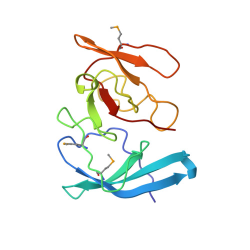
The three-dimensional structure of the RNA-binding domain of ribosomal protein L2; a protein at the peptidyl transferase center of the ribosome.

Nakagawa, A.Nakashima, T.Taniguchi, M.Hosaka, H.Kimura, M.Tanaka, I.

(1999) EMBO J 18: 1459-1467

  • DOI: https://doi.org/10.1093/emboj/18.6.1459
  • Primary Citation of Related Structures:  
    1RL2

  • PubMed Abstract: 

    Ribosomal protein L2 is the largest protein component in the ribosome. It is located at or near the peptidyl transferase center and has been a prime candidate for the peptidyl transferase activity. It binds directly to 23S rRNA and plays a crucial role in its assembly. The three-dimensional structure of the RNA-binding domain of L2 from Bacillus stearothermophilus has been determined at 2.3 A resolution by X-ray crystallography using the selenomethionyl MAD method. The RNA-binding domain of L2 consists of two recurring motifs of approximately 70 residues each. The N-terminal domain (positions 60-130) is homologous to the OB-fold, and the C-terminal domain (positions 131-201) is homologous to the SH3-like barrel. Residues Arg86 and Arg155, which have been identified by mutation experiments to be involved in the 23S rRNA binding, are located at the gate of the interface region between the two domains. The molecular architecture suggests how this important protein has evolved from the ancient nucleic acid-binding proteins to create a 23S rRNA-binding domain in the very remote past.


  • Organizational Affiliation

    Division of Biological Sciences, Graduate School of Science, Hokkaido University, Sapporo 060-0810, Japan.


Macromolecules
Find similar proteins by:  (by identity cutoff)  |  3D Structure
Entity ID: 1
MoleculeChains Sequence LengthOrganismDetailsImage
PROTEIN (RIBOSOMAL PROTEIN L2)
A, B
137Geobacillus stearothermophilusMutation(s): 0 
UniProt
Find proteins for P04257 (Geobacillus stearothermophilus)
Explore P04257 
Go to UniProtKB:  P04257
Entity Groups  
Sequence Clusters30% Identity50% Identity70% Identity90% Identity95% Identity100% Identity
UniProt GroupP04257
Sequence Annotations
Expand
  • Reference Sequence
Small Molecules
Modified Residues  1 Unique
IDChains TypeFormula2D DiagramParent
MSE
Query on MSE
A, B
L-PEPTIDE LINKINGC5 H11 N O2 SeMET
Experimental Data & Validation

Experimental Data

  • Method: X-RAY DIFFRACTION
  • Resolution: 2.30 Å
  • R-Value Free: 0.258 
  • R-Value Work: 0.203 
  • Space Group: P 1
Unit Cell:
Length ( Å )Angle ( ˚ )
a = 28.05α = 99.58
b = 36.19β = 95.86
c = 69.71γ = 102.63
Software Package:
Software NamePurpose
SHARPphasing
SOLOMONphasing
CNSrefinement
DENZOdata reduction
CCP4data scaling

Structure Validation

View Full Validation Report


Entry History 

Deposition Data

Revision History  (Full details and data files)

  • Version 1.0: 1999-04-02
    Type: Initial release
  • Version 1.1: 2008-04-26
    Changes: Version format compliance
  • Version 1.2: 2011-07-13
    Changes: Version format compliance
  • Version 1.3: 2018-01-24
    Changes: Database references
  • Version 1.4: 2023-12-27
    Changes: Data collection, Database references, Derived calculations