1RJ2 | pdb_00001rj2

Crystal structure of the DH/PH fragment of Dbs without bound GTPase


Experimental Data Snapshot

  • Method: X-RAY DIFFRACTION
  • Resolution: 3.00 Å
  • R-Value Free: 
    0.296 (Depositor), 0.291 (DCC) 
  • R-Value Work: 
    0.239 (Depositor), 0.238 (DCC) 

Starting Model: experimental
View more details

wwPDB Validation 3D Report Full Report

Validation slider image for 1RJ2

This is version 1.3 of the entry. See complete history

Literature

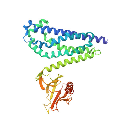
Crystal structure of the DH/PH fragment of Dbs without bound GTPase.

Worthylake, D.K.Rossman, K.L.Sondek, J.

(2004) Structure 12: 1078-1086

  • DOI: https://doi.org/10.1016/j.str.2004.03.021
  • Primary Citation Related Structures: 
    1RJ2

  • PubMed Abstract: 

    Dbl proteins are guanine nucleotide exchange factors for Rho GTPases, containing adjacent Dbl homology (DH) and pleckstrin homology (PH) domains. This domain architecture is virtually invariant and typically required for full exchange potential. Several structures of DH/PH fragments bound to GTPases implicate the PH domain in nucleotide exchange. To more fully understand the functional linkage between DH and PH domains, we have determined the crystal structure of the DH/PH fragment of Dbs without bound GTPase. This structure is generally similar to previously determined structures of Dbs bound to GTPases albeit with greater apparent mobility between the DH and PH domains. These comparisons suggest that the DH and PH domains of Dbs are spatially primed for binding GTPases and small alterations in intradomain conformations that may be elicited by subtle biological responses, such as altered phosphoinositide levels, are sufficient to enhance exchange by facilitating interactions between the PH domain and GTPases.


  • Organizational Affiliation
    • Department of Pharmacology, University of North Carolina School of Medicine, Chapel Hill, NC 27599, USA.

Macromolecule Content 

  • Total Structure Weight: 164.88 kDa 
  • Atom Count: 10,370 
  • Modeled Residue Count: 1,273 
  • Deposited Residue Count: 1,412 
  • Unique protein chains: 1

Macromolecules

Find similar proteins by:|  3D Structure
Entity ID: 1
MoleculeChains  Sequence LengthOrganismDetailsImage
Guanine nucleotide exchange factor DBS [Fragment]A,
B [auth D],
C [auth G],
D [auth J]
353Mus musculusMutation(s): 0 
Gene Names: MCF2LOST
UniProt & NIH Common Fund Data Resources
Find proteins for Q64096 (Mus musculus)
Explore Q64096 
Go to UniProtKB:  Q64096
IMPC:  MGI:103263
Entity Groups
Sequence Clusters30% Identity50% Identity70% Identity90% Identity95% Identity100% Identity
UniProt GroupQ64096
Sequence Annotations
Expand
Reference Sequence

Experimental Data & Validation

Experimental Data

  • Method: X-RAY DIFFRACTION
  • Resolution: 3.00 Å
  • R-Value Free:  0.296 (Depositor), 0.291 (DCC) 
  • R-Value Work:  0.239 (Depositor), 0.238 (DCC) 
Space Group: P 1 21 1
Unit Cell:
Length ( Å )Angle ( ˚ )
a = 97.989α = 90
b = 82.824β = 90.8
c = 127.257γ = 90
Software Package:
Software NamePurpose
DENZOdata reduction
SCALEPACKdata scaling
BEASTmodel building
CNSrefinement
BEASTphasing

Structure Validation

View Full Validation Report


Entry History 

Deposition Data

Revision History  (Full details and data files)

  • Version 1.0: 2004-06-29
    Type: Initial release
  • Version 1.1: 2008-04-29
    Changes: Version format compliance
  • Version 1.2: 2011-07-13
    Changes: Version format compliance
  • Version 1.3: 2023-08-23
    Changes: Data collection, Database references, Refinement description