Loop and subdomain movements in the mechanism of Escherichia coli dihydrofolate reductase: crystallographic evidence.
Sawaya, M.R., Kraut, J.(1997) Biochemistry 36: 586-603
- PubMed: 9012674 Search on PubMed
- DOI: https://doi.org/10.1021/bi962337c
- Primary Citation Related Structures:
1DRE, 1RA1, 1RA2, 1RA3, 1RA8, 1RA9, 1RB2, 1RB3, 1RC4, 1RD7, 1RE7, 1RF7, 1RG7, 1RH3, 1RX1, 1RX2, 1RX3, 1RX4, 1RX5, 1RX6, 1RX7, 1RX8, 1RX9 - PubMed Abstract:
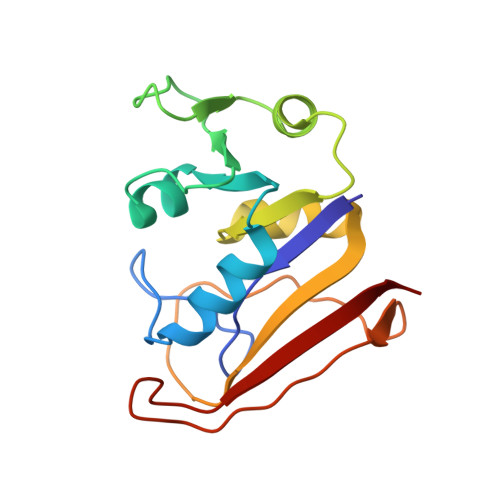
The reaction catalyzed by Escherichia coli dihydrofolate reductase (ecDHFR) cycles through five detectable kinetic intermediates: holoenzyme, Michaelis complex, ternary product complex, tetrahydrofolate (THF) binary complex, and THF.NADPH complex. Isomorphous crystal structures analogous to these five intermediates and to the transition state (as represented by the methotrexate-NADPH complex) have been used to assemble a 2.1 A resolution movie depicting loop and subdomain movements during the catalytic cycle (see Supporting Information). The structures suggest that the M20 loop is predominantly closed over the reactants in the holoenzyme, Michaelis, and transition state complexes. But, during the remainder of the cycle, when nicotinamide is not bound, the loop occludes (protrudes into) the nicotinamide-ribose binding pocket. Upon changing from the closed to the occluded conformation, the central portion of the loop rearranges from beta-sheet to 3(10) helix. The change may occur by way of an irregularly structured open loop conformation, which could transiently admit a water molecule into position to protonate N5 of dihydrofolate. From the Michaelis to the transition state analogue complex, rotation between two halves of ecDHFR, the adenosine binding subdomain and loop subdomain, closes the (p-aminobenzoyl)glutamate (pABG) binding crevice by approximately 0.5 A. Resulting enhancement of contacts with the pABG moiety may stabilize puckering at C6 of the pteridine ring in the transition state. The subdomain rotation is further adjusted by cofactor-induced movements (approximately 0.5 A) of helices B and C, producing a larger pABG cleft in the THF.NADPH analogue complex than in the THF analogue complex. Such movements may explain how THF release is assisted by NADPH binding. Subdomain rotation is not observed in vertebrate DHFR structures, but an analogous loop movement (residues 59-70) appears to similarly adjust the pABG cleft width, suggesting that these movements are important for catalysis. Loop movement, also unobserved in vertebrate DHFR structures, may preferentially weaken NADP+ vs NADPH binding in ecDHFR, an evolutionary adaptation to reduce product inhibition in the NADP+ rich environment of prokaryotes.
- Department of Chemistry and Biochemistry, University of California, San Diego, La Jolla 92093-0506, USA.
Organizational Affiliation: