1RF5 | pdb_00001rf5

Structural Studies of Streptococcus pneumoniae EPSP Synthase in Unliganded State


Experimental Data Snapshot

  • Method: X-RAY DIFFRACTION
  • Resolution: 2.30 Å
  • R-Value Free: 
    0.277 (Depositor), 0.325 (DCC) 
  • R-Value Work: 
    0.218 (Depositor), 0.312 (DCC) 
  • R-Value Observed: 
    0.221 (Depositor) 

wwPDB Validation 3D Report Full Report

Validation slider image for 1RF5

This is version 1.3 of the entry. See complete history

Literature

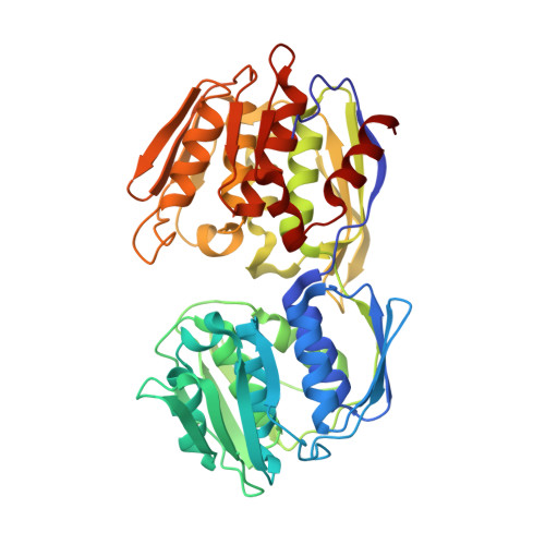
Structural studies of Streptococcus pneumoniae EPSP synthase in unliganded state, tetrahedral intermediate-bound state and S3P-GLP-bound state.

Park, H.Hilsenbeck, J.L.Kim, H.J.Shuttleworth, W.A.Park, Y.H.Evans, J.N.Kang, C.

(2004) Mol Microbiol 51: 963-971

  • DOI: https://doi.org/10.1046/j.1365-2958.2003.03885.x
  • Primary Citation Related Structures: 
    1RF4, 1RF5, 1RF6

  • PubMed Abstract: 

    The shikimate pathway synthesizes aromatic amino acids and other essential metabolites that are necessary for bacteria, plants and fungi to survive. This pathway is not present in vertebrates and therefore represents an attractive target for antibacterial agents. We have successfully crystallized and solved the structure of unliganded, inhibitor-liganded and tetrahedral intermediate (TI)-liganded forms of Streptococcus pneumoniae EPSP synthase. The overall topology of the S. pneumoniae EPSP synthase is similar to that of the Escherichia coli EPSP synthase. In addition, the majority of residues responsible for ligand binding were conserved between the two proteins. TI-liganded structure provides absolute configuration of the C-2 atom from the F-PEP moiety of the enzyme-bound intermediate and also defines key residues responsible for the enzyme reaction. Comparison of the unliganded state and substrate-bound state of the enzyme provides insights into the structural mechanisms involved in dynamic events of ligand binding, domain movement and closure. This structural study of the pathogenic bacteria S. pneumoniae EPSP synthase with inhibitor and TI will provide invaluable information for the design of new-generation antibiotics.


  • Organizational Affiliation
    • School of Molecular Biosciences, Washington State University, Pullman 99164, USA.

Macromolecule Content 

  • Total Structure Weight: 183.52 kDa 
  • Atom Count: 13,615 
  • Modeled Residue Count: 1,708 
  • Deposited Residue Count: 1,708 
  • Unique protein chains: 1

Macromolecules

Find similar proteins by:|  3D Structure
Entity ID: 1
MoleculeChains  Sequence LengthOrganismDetailsImage
5-enolpyruvylshikimate-3-phosphate synthase
A, B, C, D
427Streptococcus pneumoniaeMutation(s): 0 
Gene Names: aroA
EC: 2.5.1.19
UniProt
Find proteins for Q9S400 (Streptococcus pneumoniae serotype 4 (strain ATCC BAA-334 / TIGR4))
Explore Q9S400 
Go to UniProtKB:  Q9S400
Entity Groups
Sequence Clusters30% Identity50% Identity70% Identity90% Identity95% Identity100% Identity
UniProt GroupQ9S400
Sequence Annotations
Expand
Reference Sequence

Experimental Data & Validation

Experimental Data

  • Method: X-RAY DIFFRACTION
  • Resolution: 2.30 Å
  • R-Value Free:  0.277 (Depositor), 0.325 (DCC) 
  • R-Value Work:  0.218 (Depositor), 0.312 (DCC) 
  • R-Value Observed: 0.221 (Depositor) 
Space Group: P 21 21 21
Unit Cell:
Length ( Å )Angle ( ˚ )
a = 115.592α = 90
b = 116.482β = 90
c = 176.323γ = 90
Software Package:
Software NamePurpose
Blu-Icedata collection
SCALEPACKdata scaling
SOLVEphasing
X-PLORrefinement

Structure Validation

View Full Validation Report


Entry History 

Deposition Data

Revision History  (Full details and data files)

  • Version 1.0: 2004-02-17
    Type: Initial release
  • Version 1.1: 2008-04-29
    Changes: Version format compliance
  • Version 1.2: 2011-07-13
    Changes: Version format compliance
  • Version 1.3: 2024-02-14
    Changes: Data collection, Database references