1REU | pdb_00001reu

Structure of the bone morphogenetic protein 2 mutant L51P


Experimental Data Snapshot

  • Method: X-RAY DIFFRACTION
  • Resolution: 2.65 Å
  • R-Value Free: 
    0.235 (Depositor), 0.238 (DCC) 
  • R-Value Work: 
    0.215 (Depositor), 0.213 (DCC) 
  • R-Value Observed: 
    0.215 (Depositor) 

Starting Model: experimental
View more details

wwPDB Validation 3D Report Full Report

Validation slider image for 1REU

This is version 1.5 of the entry. See complete history

Literature

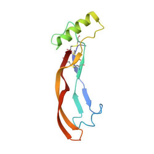
Molecular recognition of BMP-2 and BMP receptor IA.

Keller, S.Nickel, J.Zhang, J.L.Sebald, W.Mueller, T.D.

(2004) Nat Struct Mol Biol 11: 481-488

  • DOI: https://doi.org/10.1038/nsmb756
  • Primary Citation Related Structures: 
    1REU, 1REW

  • PubMed Abstract: 

    Bone morphogenetic protein-2 (BMP-2) and other members of the TGF-beta superfamily regulate the development, maintenance and regeneration of tissues and organs. Binding epitopes for these extracellular signaling proteins have been defined, but hot spots specifying binding affinity and specificity have so far not been identified. In this study, mutational and structural analyses show that epitopes of BMP-2 and the BRIA receptor form a new type of protein-protein interface. The main chain atoms of Leu 51 and Asp53 of BMP-2 represent a hot spot of binding to BRIA. The BMP-2 variant L51P was deficient in type I receptor binding only, whereas its overall structure and its binding to type II receptors and modulator proteins, such as noggin, were unchanged. Thus, the L51P substitution converts BMP-2 into a receptor-inactive inhibitor of noggin. These results are relevant for other proteins of the TGF-beta superfamily and provide useful clues for structure-based drug design.


  • Organizational Affiliation
    • Lehrstuhl für Physiologische Chemie II, Theodor-Boveri Institut für Biowissenschaften (Biozentrum), Universität Würzburg, Am Hubland, D-97074 Wuerzburg, Germany.

Macromolecule Content 

  • Total Structure Weight: 11.73 kDa 
  • Atom Count: 833 
  • Modeled Residue Count: 103 
  • Deposited Residue Count: 103 
  • Unique protein chains: 1

Macromolecules

Find similar proteins by:|  3D Structure
Entity ID: 1
MoleculeChains  Sequence LengthOrganismDetailsImage
bone morphogenetic protein 2103Homo sapiensMutation(s): 1 
UniProt & NIH Common Fund Data Resources
Find proteins for P12643 (Homo sapiens)
Explore P12643 
Go to UniProtKB:  P12643
PHAROS:  P12643
GTEx:  ENSG00000125845 
Entity Groups
Sequence Clusters30% Identity50% Identity70% Identity90% Identity95% Identity100% Identity
UniProt GroupP12643
Sequence Annotations
Expand
Reference Sequence

Experimental Data & Validation

Experimental Data

  • Method: X-RAY DIFFRACTION
  • Resolution: 2.65 Å
  • R-Value Free:  0.235 (Depositor), 0.238 (DCC) 
  • R-Value Work:  0.215 (Depositor), 0.213 (DCC) 
  • R-Value Observed: 0.215 (Depositor) 
Space Group: H 3 2
Unit Cell:
Length ( Å )Angle ( ˚ )
a = 94.292α = 90
b = 94.292β = 90
c = 102.882γ = 120
Software Package:
Software NamePurpose
MOSFLMdata reduction
SCALAdata scaling
CNSrefinement
CCP4data scaling
CNSphasing

Structure Validation

View Full Validation Report


Entry History 

Deposition Data

Revision History  (Full details and data files)

  • Version 1.0: 2004-05-04
    Type: Initial release
  • Version 1.1: 2008-04-29
    Changes: Version format compliance
  • Version 1.2: 2011-07-13
    Changes: Derived calculations, Version format compliance
  • Version 1.3: 2021-10-27
    Changes: Database references, Derived calculations
  • Version 1.4: 2023-08-23
    Changes: Data collection, Refinement description
  • Version 1.5: 2024-10-09
    Changes: Structure summary