1RDA | pdb_00001rda

CRYSTAL STRUCTURES OF RIBONUCLEASE HI ACTIVE SITE MUTANTS FROM ESCHERICHIA COLI


Experimental Data Snapshot

  • Method: X-RAY DIFFRACTION
  • Resolution: 2.15 Å
  • R-Value Work: 
    0.180 (DCC) 
  • R-Value Observed: 
    0.189 (Depositor) 

wwPDB Validation 3D Report Full Report

Validation slider image for 1RDA

This is version 1.4 of the entry. See complete history

Literature

Crystal structures of ribonuclease HI active site mutants from Escherichia coli.

Katayanagi, K.Ishikawa, M.Okumura, M.Ariyoshi, M.Kanaya, S.Kawano, Y.Suzuki, M.Tanaka, I.Morikawa, K.

(1993) J Biological Chem 268: 22092-22099

  • Primary Citation Related Structures: 
    1RDA, 1RDB, 1RDC

  • PubMed Abstract: 

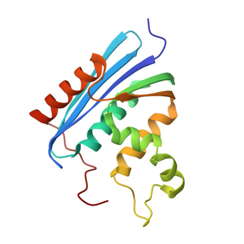
    In order to investigate the relationships between the three-dimensional structure and the enzymic activity of E. coli RNase HI, three mutant proteins, which were completely inactivated by the replacements of three functional residues, Asp10 by Asn (D10N), Glu48 by Gln (E48Q), and Asp70 by Asn (D70N), were crystallized. Their three-dimensional structures were determined by x-ray crystallography. Although the entire backbone structures of these mutants were not affected by the replacements, very localized conformational changes were observed around the Mg(2+)-binding site. The substitution of an amide group for a negatively charged carboxyl group in common induces the formation of new hydrogen bond networks, presumably due to the cancellation of repulsive forces between carboxyl side chains with negative charges. These conformational changes can account for the loss of the enzymic activity in the mutants, and suggest a possible role for Mg2+ in the hydrolysis. Since the 3 replaced acidic residues are completely conserved in sequences of reverse transcriptases from retroviruses, including human immunodeficiency virus, the concepts of the catalytic mechanism deduced from this structural analysis can also be applied to RNase H activity in reverse transcriptases.


  • Organizational Affiliation
    • Protein Engineering Research Institute, Osaka, Japan.

Macromolecule Content 

  • Total Structure Weight: 17.62 kDa 
  • Atom Count: 1,338 
  • Modeled Residue Count: 155 
  • Deposited Residue Count: 155 
  • Unique protein chains: 1

Macromolecules

Find similar proteins by:|  3D Structure
Entity ID: 1
MoleculeChains  Sequence LengthOrganismDetailsImage
RIBONUCLEASE H155Escherichia coliMutation(s): 0 
EC: 3.1.26.4
UniProt
Find proteins for P0A7Y4 (Escherichia coli (strain K12))
Explore P0A7Y4 
Go to UniProtKB:  P0A7Y4
Entity Groups
Sequence Clusters30% Identity50% Identity70% Identity90% Identity95% Identity100% Identity
UniProt GroupP0A7Y4
Sequence Annotations
Expand
Reference Sequence

Experimental Data & Validation

Experimental Data

  • Method: X-RAY DIFFRACTION
  • Resolution: 2.15 Å
  • R-Value Work:  0.180 (DCC) 
  • R-Value Observed: 0.189 (Depositor) 
Space Group: P 21 21 21
Unit Cell:
Length ( Å )Angle ( ˚ )
a = 44.17α = 90
b = 87.23β = 90
c = 35.27γ = 90
Software Package:
Software NamePurpose
PROLSQrefinement

Structure Validation

View Full Validation Report


Entry History 

Deposition Data

Revision History  (Full details and data files)

  • Version 1.0: 1993-10-31
    Type: Initial release
  • Version 1.1: 2008-03-24
    Changes: Version format compliance
  • Version 1.2: 2011-07-13
    Changes: Version format compliance
  • Version 1.3: 2017-11-29
    Changes: Derived calculations, Other
  • Version 1.4: 2024-02-14
    Changes: Data collection, Database references