1RD6 | pdb_00001rd6

Crystal Structure of S. Marcescens Chitinase A Mutant W167A


Experimental Data Snapshot

  • Method: X-RAY DIFFRACTION
  • Resolution: 2.60 Å
  • R-Value Free: 
    0.235 (Depositor) 
  • R-Value Work: 
    0.195 (Depositor), 0.209 (DCC) 
  • R-Value Observed: 
    0.195 (Depositor) 

Starting Model: experimental
View more details

wwPDB Validation 3D Report Full Report

Validation slider image for 1RD6

This is version 1.5 of the entry. See complete history

Literature

Mutation of a conserved tryptophan in the chitin-binding cleft of Serratia marcescens chitinase A enhances transglycosylation.

Aronson, N.N.Halloran, B.A.Alexeyev, M.F.Zhou, X.E.Wang, Y.Meehan, E.J.Chen, L.

(2006) Biosci Biotechnol Biochem 70: 243-251

  • DOI: https://doi.org/10.1271/bbb.70.243
  • Primary Citation Related Structures: 
    1RD6

  • PubMed Abstract: 

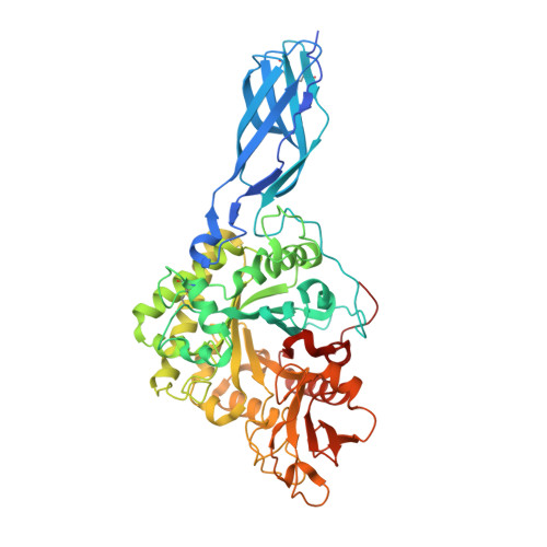
    Family 18 chitinases have the signature peptide DGXDXDXE forming the fourth beta-strand in the (beta/alpha)8-barrel of their catalytic domain. The carboxyl-end glutamic acid, E315 in Serratia marcescens chitinase A, serves as the acid/base during chitin hydrolysis, and the side-chain of the preceding aspartic acid, D313, helps to position correctly the N-acetyl moiety of the glycosyl sugar undergoing hydrolysis. Chitin substrates are bound within a long cleft across the top of the barrel, whose floor consists of aromatic residues that hydrophobically stack with every other GlcNAc. Alanine substitution of the conserved Trp167 at the -3 subsite in Serratia marcescens chitinase A enhanced transglycosylation. Higher oligosaccharides were formed from both chitin tetra- and pentasaccharide, and the only hydrolytic product from chitin trisaccharide was the disaccharide. Greater retention of the glycosyl fragment at the active site of the -3 mutant of Serratia marcescens chitinase A might favor transglycosylation due to a stabilized conformation of its D313.


  • Organizational Affiliation
    • Department of Biochemistry and Molecular Biology, University of South Alabama, AL 36688, USA. naronson@jaguar1.usouthal.edu

Macromolecule Content 

  • Total Structure Weight: 60.97 kDa 
  • Atom Count: 4,413 
  • Modeled Residue Count: 540 
  • Deposited Residue Count: 563 
  • Unique protein chains: 1

Macromolecules

Find similar proteins by:|  3D Structure
Entity ID: 1
MoleculeChains  Sequence LengthOrganismDetailsImage
Chitinase A563Serratia marcescensMutation(s): 1 
Gene Names: CHIA
EC: 3.2.1.14
UniProt
Find proteins for P07254 (Serratia marcescens)
Explore P07254 
Go to UniProtKB:  P07254
Entity Groups
Sequence Clusters30% Identity50% Identity70% Identity90% Identity95% Identity100% Identity
UniProt GroupP07254
Sequence Annotations
Expand
Reference Sequence

Experimental Data & Validation

Experimental Data

  • Method: X-RAY DIFFRACTION
  • Resolution: 2.60 Å
  • R-Value Free:  0.235 (Depositor) 
  • R-Value Work:  0.195 (Depositor), 0.209 (DCC) 
  • R-Value Observed: 0.195 (Depositor) 
Space Group: I 2 2 2
Unit Cell:
Length ( Å )Angle ( ˚ )
a = 77.588α = 90
b = 134.065β = 90
c = 187.904γ = 90
Software Package:
Software NamePurpose
CNSrefinement
HKL-2000data reduction
SCALEPACKdata scaling
CNSphasing

Structure Validation

View Full Validation Report


Entry History 

Deposition Data

Revision History  (Full details and data files)

  • Version 1.0: 2004-12-07
    Type: Initial release
  • Version 1.1: 2008-04-29
    Changes: Version format compliance
  • Version 1.2: 2011-07-13
    Changes: Version format compliance
  • Version 1.3: 2021-10-27
    Changes: Database references
  • Version 1.4: 2023-08-23
    Changes: Data collection, Refinement description
  • Version 1.5: 2024-11-20
    Changes: Structure summary