1RAS | pdb_00001ras

CRYSTAL STRUCTURE OF A FLUORESCENT DERIVATIVE OF RNASE A


Experimental Data Snapshot

  • Method: X-RAY DIFFRACTION
  • Resolution: 1.70 Å
  • R-Value Observed: 
    0.203 (Depositor) 

wwPDB Validation 3D Report Full Report

Validation slider image for 1RAS

This is version 1.4 of the entry. See complete history

Literature

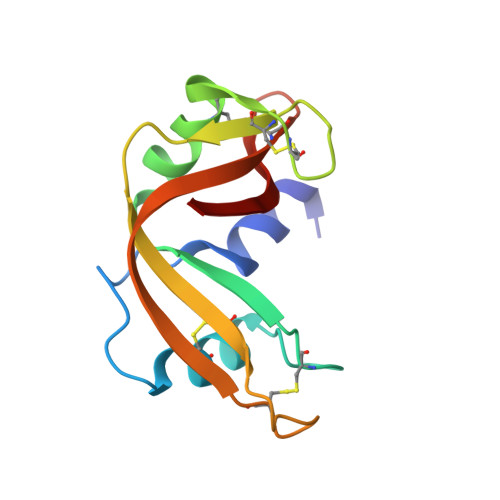
Crystal structure of a fluorescent derivative of RNase A.

Baudet-Nessler, S.Jullien, M.Crosio, M.P.Janin, J.

(1993) Biochemistry 32: 8457-8464

  • Primary Citation Related Structures: 
    1RAR, 1RAS

  • PubMed Abstract: 

    The crystal structure of RNase A chemically modified with the fluorescent probe, N-[[(iodoacetyl)-amino]ethyl]-5-naphthylamine-1-sulfonic acid (1,5-IAENS), has been solved and refined to high resolution. It yields information on the mode of binding, the mobility of a probe commonly used in spectroscopic studies, and anion binding sites in RNase A. Trigonal crystals of the fluorescent derivative grown in sodium or cesium chloride and ammonium sulfate, pH 5.1, were nearly isomorphous with those of a semisynthetic RNase [DeMel, et al. (1992) J. Biol. Chem. 267, 247-256]. Refinement starting from semisynthetic RNase led to a model with R = 20% against 1.7-A diffraction data from crystals in ammonium sulfate and another model with R = 17% against 1.9-A data taken in the presence of 3 M NaCl. The second model contains three chloride ions: one is at the active site, and the other two are at molecular interfaces. Otherwise, the two models are very similar. The fluorophore has very little effect on the protein conformation. It is found to be covalently attached to the active site His-12 with the naphthyl group stacked on the imidazole ring of His-119. It remains largely accessible to solvent and in a polar environment on the protein surface, even though the fluorescence emission spectrum is blue shifted as it is in nonpolar solvents.


  • Organizational Affiliation
    • Laboratoire de Biologie Structurale, UMR 9920 CNRS-Université Paris-Sud, Orsay, France.

Macromolecule Content 

  • Total Structure Weight: 13.89 kDa 
  • Atom Count: 1,063 
  • Modeled Residue Count: 123 
  • Deposited Residue Count: 123 
  • Unique protein chains: 1

Macromolecules

Find similar proteins by:|  3D Structure
Entity ID: 1
MoleculeChains  Sequence LengthOrganismDetailsImage
RIBONUCLEASE A123Bos taurusMutation(s): 0 
EC: 3.1.27.5 (PDB Primary Data), 4.6.1.18 (UniProt)
UniProt
Find proteins for P61823 (Bos taurus)
Explore P61823 
Go to UniProtKB:  P61823
Entity Groups
Sequence Clusters30% Identity50% Identity70% Identity90% Identity95% Identity100% Identity
UniProt GroupP61823
Sequence Annotations
Expand
Reference Sequence

Small Molecules

Ligands 1 Unique
IDChains Name / Formula / InChI Key2D Diagram3D Interactions
AEN

Query on AEN


Download:Ideal Coordinates CCD File
B [auth A]5-(1-SULFONAPHTHYL)-ACETYLAMINO-ETHYLAMINE
C14 H16 N2 O4 S
FBZFLXJHAMMUQM-UHFFFAOYSA-N

Experimental Data & Validation

Experimental Data

  • Method: X-RAY DIFFRACTION
  • Resolution: 1.70 Å
  • R-Value Observed: 0.203 (Depositor) 
Space Group: P 32 2 1
Unit Cell:
Length ( Å )Angle ( ˚ )
a = 65.1α = 90
b = 65.1β = 90
c = 65.02γ = 120
Software Package:
Software NamePurpose
PROLSQrefinement

Structure Validation

View Full Validation Report


Entry History 

Deposition Data

Revision History  (Full details and data files)

  • Version 1.0: 1993-10-31
    Type: Initial release
  • Version 1.1: 2008-03-24
    Changes: Version format compliance
  • Version 1.2: 2011-07-13
    Changes: Version format compliance
  • Version 1.3: 2024-06-05
    Changes: Data collection, Database references, Derived calculations, Other
  • Version 1.4: 2024-11-20
    Changes: Structure summary