Three-dimensional structure of rat liver 3 alpha-hydroxysteroid/dihydrodiol dehydrogenase: a member of the aldo-keto reductase superfamily.
Hoog, S.S., Pawlowski, J.E., Alzari, P.M., Penning, T.M., Lewis, M.(1994) Proc Natl Acad Sci U S A 91: 2517-2521
- PubMed: 8146147 Search on PubMedSearch on PubMed Central
- DOI: https://doi.org/10.1073/pnas.91.7.2517
- Primary Citation Related Structures:
1RAL - PubMed Abstract:
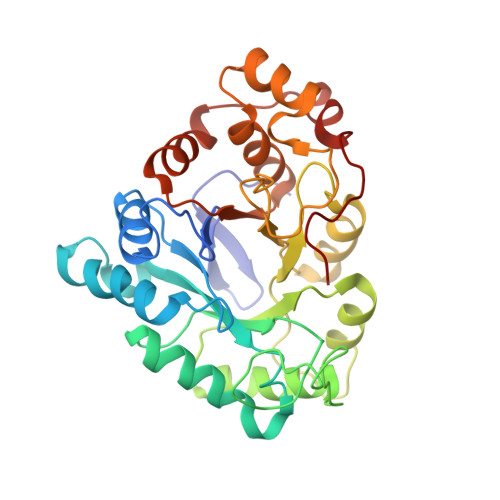
The 3.0-A-resolution x-ray structure of rat liver 3 alpha-hydroxysteroid dehydrogenase/dihydrodiol dehydrogenase (3 alpha-HSD, EC 1.1.1.50) was determined by molecular replacement using human placental aldose reductase as the search model. The protein folds into an alpha/beta or triose-phosphate isomerase barrel and lacks a canonical Rossmann fold for binding pyridine nucleotide. The structure contains a concentration of hydrophobic amino acids that lie in a cavity near the top of the barrel and that are presumed to be involved in binding hydrophobic substrates (steroids, prostaglandins, and polycyclic aromatic hydrocarbons) and inhibitors (nonsteroidal antiinflammatory drugs). At the distal end of this cavity lie three residues in close proximity that have been implicated in catalysis by site-directed mutagenesis--Tyr-55, Asp-50, and Lys-84. Tyr-55 is postulated to act as the general acid. 3 alpha-HSD shares significant sequence identity with other HSDs that belong to the aldo-keto reductase superfamily and these may show similar architecture. Other members of this family include prostaglandin F synthase and rho-crystallin. By contrast, 3 alpha-HSD shares no sequence identity with HSDs that are members of the short-chain alcohol dehydrogenase family but does contain the Tyr-Xaa-Xaa-Xaa-Lys consensus sequence implicated in catalysis in this family. In the 3 alpha-HSD structure these residues are on the periphery of the barrel and are unlikely to participate in catalysis.
- Department of Biochemistry and Biophysics, University of Pennsylvania School of Medicine, Philadelphia 19104-6084.
Organizational Affiliation: