1R6J | pdb_00001r6j

Ultrahigh resolution Crystal Structure of syntenin PDZ2


Experimental Data Snapshot

  • Method: X-RAY DIFFRACTION
  • Resolution: 0.73 Å
  • R-Value Free: 
    0.087 (Depositor), 0.080 (DCC) 
  • R-Value Work: 
    0.075 (Depositor), 0.078 (DCC) 

wwPDB Validation 3D Report Full Report

Validation slider image for 1R6J

This is version 1.3 of the entry. See complete history

Literature

The PDZ2 domain of syntenin at ultra-high resolution: bridging the gap between small molecule and macromolecular crystal chemistry

Kang, B.S.Devedjiev, Y.Derewenda, U.Derewenda, Z.S.

(2004) J Mol Biology 338: 483-493

  • DOI: https://doi.org/10.1016/j.jmb.2004.02.057
  • Primary Citation Related Structures: 
    1R6J

  • PubMed Abstract: 

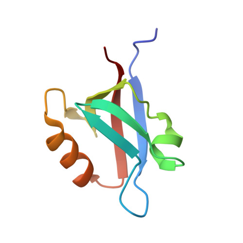
    The crystal structure of the second PDZ domain of the scaffolding protein syntenin was solved using data extending to 0.73 A resolution. The crystallographic model, including the hydrogen atoms and the anisotropic displacement parameters, was refined to a conventional R-factor of 7.5% and Rfree of 8.7%, making it the most precise crystallographic model of a protein molecule to date. The model reveals discrete disorder in several places in the molecule, and significant plasticity of the peptide bond, with some omega angles deviating by nearly 20 degrees from planarity. Most hydrogen atoms are easily identifiable in the electron density and weak hydrogen bonds of the C-H...O type are clearly visible between the beta-strands. The study sets a new standard for high-resolution protein crystallography.


  • Organizational Affiliation
    • Department of Molecular Physiology and Biological Physics, University of Virginia, Charlottesville, VA 22908-0736, USA.

Macromolecule Content 

  • Total Structure Weight: 8.75 kDa 
  • Atom Count: 964 
  • Modeled Residue Count: 82 
  • Deposited Residue Count: 82 
  • Unique protein chains: 1

Macromolecules

Find similar proteins by:|  3D Structure
Entity ID: 1
MoleculeChains  Sequence LengthOrganismDetailsImage
Syntenin 182Homo sapiensMutation(s): 0 
Gene Names: SDCBP OR MDA9 OR SYCL
UniProt & NIH Common Fund Data Resources
Find proteins for O00560 (Homo sapiens)
Explore O00560 
Go to UniProtKB:  O00560
PHAROS:  O00560
GTEx:  ENSG00000137575 
Entity Groups
Sequence Clusters30% Identity50% Identity70% Identity90% Identity95% Identity100% Identity
UniProt GroupO00560
Sequence Annotations
Expand
Reference Sequence

Small Molecules

Ligands 1 Unique
IDChains Name / Formula / InChI Key2D Diagram3D Interactions
CL

Query on CL


Download:Ideal Coordinates CCD File
B [auth A]CHLORIDE ION
Cl
VEXZGXHMUGYJMC-UHFFFAOYSA-M

Experimental Data & Validation

Experimental Data

  • Method: X-RAY DIFFRACTION
  • Resolution: 0.73 Å
  • R-Value Free:  0.087 (Depositor), 0.080 (DCC) 
  • R-Value Work:  0.075 (Depositor), 0.078 (DCC) 
Space Group: P 1 21 1
Unit Cell:
Length ( Å )Angle ( ˚ )
a = 25.876α = 90
b = 39.544β = 109.64
c = 32.276γ = 90
Software Package:
Software NamePurpose
SHELXmodel building
SHELXL-97refinement
HKL-2000data reduction
SCALEPACKdata scaling
SHELXphasing

Structure Validation

View Full Validation Report


Entry History 

Deposition Data

Revision History  (Full details and data files)

  • Version 1.0: 2004-05-04
    Type: Initial release
  • Version 1.1: 2008-04-29
    Changes: Version format compliance
  • Version 1.2: 2011-07-13
    Changes: Version format compliance
  • Version 1.3: 2024-02-14
    Changes: Data collection, Database references, Derived calculations