Role of the amino sugar in DNA binding of disaccharide anthracyclines: crystal structure of the complex MAR70/d(CGATCG).
Temperini, C., Cirilli, M., Aschi, M., Ughetto, G.(2005) Bioorg Med Chem 13: 1673-1679
- PubMed: 15698785 Search on PubMed
- DOI: https://doi.org/10.1016/j.bmc.2004.12.007
- Primary Citation Related Structures:
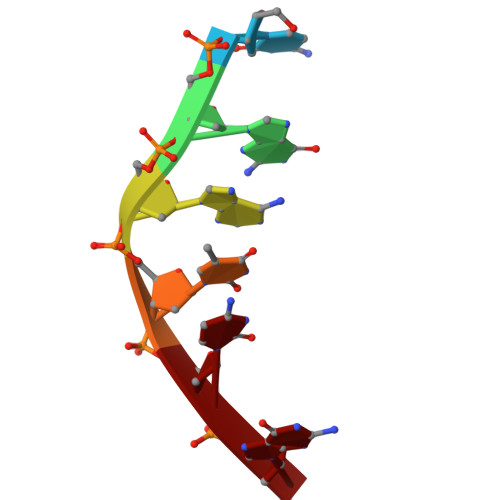
1R68 - PubMed Abstract:
Disaccharide anthracyclines analogues have been shown to exhibit different antitumour activity as compared with parents compounds doxorubicin and daunomycin. Here we report the crystal structure of the disaccharide analog MAR70 complexed with the DNA hexamer d(CGATCG). The structure has been solved at 1.54A resolution and is similar to previous crystallized anthracycline-DNA complexes with both sugar rings of the disaccharide chain lying in the DNA minor groove. Comparison with the structure of MEN10755 another disaccharide anthracycline co-crystallized with the same DNA hexamer suggests a correlation between the position of the amino sugar on the disaccharide chain and the conformation of this moiety when binding to DNA. This is discussed with respect to the influence on drug activity and on the possible interaction with other cellular targets.
- Department of Chemistry, University of Florence, 50019 Sesto F.no (FI), Italy.
Organizational Affiliation: