Combined pseudo-merohedral twinning, non-crystallographic symmetry and pseudo-translation in a monoclinic crystal form of the gammadelta T-cell ligand T10.
Rudolph, M.G., Wingren, C., Crowley, M.P., Chien, Y.H., Wilson, I.A.(2004) Acta Crystallogr D Biol Crystallogr 60: 656-664
- PubMed: 15039553 Search on PubMed
- DOI: https://doi.org/10.1107/S0907444904002239
- Primary Citation Related Structures:
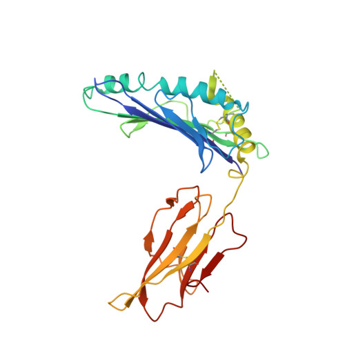
1R3H - PubMed Abstract:
T10 is a non-classical class Ib-like major histocompatibility complex (MHC) cell-surface antigen which binds directly to certain gammadelta T-cell receptors in the absence of any exogenous and endogenous ligands, such as peculiar lipids or glycolipids. The crystal structure at 2.5 A resolution of murine T10 was determined by molecular replacement using data from an almost perfectly twinned monoclinic crystal. The space group is P2(1), with unit-cell parameters a = 78.2, b = 70.0, c = 139.2 A, beta = 106.8 degrees. Self-rotation function analysis and various intensity statistics revealed the presence of pseudo-merohedral twinning, but these tests underestimated the true twin fraction of alpha approximately 0.46. Native Patterson analyses pointed to the presence of pseudo-translation among the four molecules present in the asymmetric unit. Data analysis, structure determination and model refinement are discussed.
- Department of Molecular Biology and The Skaggs Institute for Chemical Biology, The Scripps Research Institute, La Jolla, CA 92037, USA. markus.rudolph@bio.uni-goettingen.de
Organizational Affiliation: