1R1X | pdb_00001r1x

Crystal structure of oxy-human hemoglobin Bassett at 2.15 angstrom


Experimental Data Snapshot

  • Method: X-RAY DIFFRACTION
  • Resolution: 2.15 Å
  • R-Value Free: 
    0.288 (Depositor), 0.277 (DCC) 
  • R-Value Work: 
    0.222 (Depositor), 0.215 (DCC) 
  • R-Value Observed: 
    0.222 (Depositor) 

Starting Model: experimental
View more details

wwPDB Validation 3D Report Full Report

Validation slider image for 1R1X

Ligand Structure Quality Assessment 


This is version 1.5 of the entry. See complete history

Literature

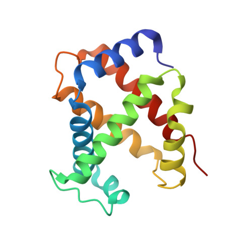
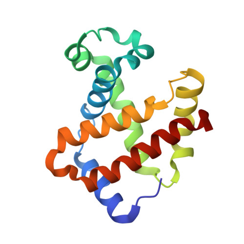
Characterization of hemoglobin bassett (alpha94Asp-->Ala), a variant with very low oxygen affinity

Abdulmalik, O.Safo, M.K.Lerner, N.B.Ochotorena, J.Daikhin, Y.Lakka, V.Santacroce, R.Abraham, D.J.Asakura, T.

(2004) Am J Hematol 77: 268-276

  • DOI: https://doi.org/10.1002/ajh.20184
  • Primary Citation Related Structures: 
    1R1X, 1R1Y

  • PubMed Abstract: 

    Hemoglobin (Hb) Bassett, an abnormal Hb variant with a markedly reduced oxygen affinity, was discovered in a Caucasian (Anglo-Saxon) male child who experienced episodes of cyanosis. Cation-exchange and reversed-phase (RP) high-performance liquid chromatography (HPLC) showed that the patient has an abnormal Hb, with a mutation in the alpha-globin. Tryptic peptide digest of the abnormal alpha-globin with subsequent HPLC analysis revealed abnormal elution of the alpha-T11 peptide. Further studies with Edman sequencing and electrospray mass spectrometry of tryptic peptide alpha-T11, as well as structural analysis by X-ray crystallography revealed an Asp-->Ala substitution at the alpha94 (G1) position, a match for Hb Bassett. Detailed functional studies showed that this Hb variant had a markedly reduced oxygen affinity (P(50) at pH 7.0 = 22 mmHg; Hb A P(50) = 10.5 mmHg), reduced Bohr effect (-0.26 compared to - 0.54 in Hb A), and low subunit cooperativity (n = 1.4, compared to 2.6 in Hb A). X-ray crystallography results explain the probable effects of the structural modification on the oxygen-binding properties of this Hb variant.


  • Organizational Affiliation
    • Division of Hematology, The Children's Hospital of Philadelphia, Philadelphia, Pennsylvania 19104, USA.

Macromolecule Content 

  • Total Structure Weight: 32.47 kDa 
  • Atom Count: 2,485 
  • Modeled Residue Count: 287 
  • Deposited Residue Count: 287 
  • Unique protein chains: 2

Macromolecules

Find similar proteins by:|  3D Structure
Entity ID: 1
MoleculeChains  Sequence LengthOrganismDetailsImage
Hemoglobin alpha chain141Homo sapiensMutation(s): 1 
UniProt & NIH Common Fund Data Resources
Find proteins for P69905 (Homo sapiens)
Explore P69905 
Go to UniProtKB:  P69905
Entity Groups
Sequence Clusters30% Identity50% Identity70% Identity90% Identity95% Identity100% Identity
UniProt GroupP69905
Sequence Annotations
Expand
Reference Sequence
Find similar proteins by:|  3D Structure
Entity ID: 2
MoleculeChains  Sequence LengthOrganismDetailsImage
Hemoglobin beta chain146Homo sapiensMutation(s): 0 
UniProt & NIH Common Fund Data Resources
Find proteins for P68871 (Homo sapiens)
Explore P68871 
Go to UniProtKB:  P68871
PHAROS:  P68871
GTEx:  ENSG00000244734 
Entity Groups
Sequence Clusters30% Identity50% Identity70% Identity90% Identity95% Identity100% Identity
UniProt GroupP68871
Sequence Annotations
Expand
Reference Sequence

Experimental Data & Validation

Experimental Data

  • Method: X-RAY DIFFRACTION
  • Resolution: 2.15 Å
  • R-Value Free:  0.288 (Depositor), 0.277 (DCC) 
  • R-Value Work:  0.222 (Depositor), 0.215 (DCC) 
  • R-Value Observed: 0.222 (Depositor) 
Space Group: P 41 21 2
Unit Cell:
Length ( Å )Angle ( ˚ )
a = 53.357α = 90
b = 53.357β = 90
c = 191.781γ = 90
Software Package:
Software NamePurpose
CNSrefinement
bioteXdata reduction
bioteXdata scaling
CNSphasing

Structure Validation

View Full Validation Report


Ligand Structure Quality Assessment 


Entry History 

Deposition Data

Revision History  (Full details and data files)

  • Version 1.0: 2003-10-07
    Type: Initial release
  • Version 1.1: 2008-04-29
    Changes: Version format compliance
  • Version 1.2: 2011-07-13
    Changes: Version format compliance
  • Version 1.3: 2017-10-11
    Changes: Refinement description
  • Version 1.4: 2018-08-29
    Changes: Data collection, Database references
  • Version 1.5: 2023-08-23
    Changes: Data collection, Database references, Derived calculations, Refinement description