1R1I | pdb_00001r1i

STRUCTURAL ANALYSIS OF NEPRILYSIN WITH VARIOUS SPECIFIC AND POTENT INHIBITORS


Experimental Data Snapshot

  • Method: X-RAY DIFFRACTION
  • Resolution: 2.60 Å
  • R-Value Free: 
    0.358 (Depositor), 0.331 (DCC) 
  • R-Value Work: 
    0.276 (Depositor), 0.262 (DCC) 
  • R-Value Observed: 
    0.280 (Depositor) 

wwPDB Validation 3D Report Full Report

Validation slider image for 1R1I

Ligand Structure Quality Assessment 


This is version 1.4 of the entry. See complete history

Literature

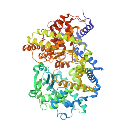
Structural analysis of neprilysin with various specific and potent inhibitors.

Oefner, C.Roques, B.P.Fournie-Zaluski, M.C.Dale, G.E.

(2004) Acta Crystallogr D Biol Crystallogr 60: 392-396

  • DOI: https://doi.org/10.1107/S0907444903027410
  • Primary Citation Related Structures: 
    1R1H, 1R1I, 1R1J

  • PubMed Abstract: 

    Neutral endopeptidase (NEP) is the major enzyme involved in the metabolic inactivation of a number of bioactive peptides including the enkephalins, substance P, endothelin, bradykinin and atrial natriuretic factor. Owing to the physiological importance of NEP in the modulation of nociceptive and pressor responses, there is considerable interest in inhibitors of this enzyme as novel analgesics and antihypertensive agents. Here, the crystal structures of the soluble extracellular domain of human NEP (residues 52-749) complexed with various potent and competitive inhibitors are described. The structures unambiguously reveal the binding mode of the different zinc-chelating groups and the subsite specificity of the enzyme.


  • Organizational Affiliation
    • Morphochem AG, CH-4058 Basel, Switzerland.

Macromolecule Content 

  • Total Structure Weight: 80.64 kDa 
  • Atom Count: 5,683 
  • Modeled Residue Count: 696 
  • Deposited Residue Count: 696 
  • Unique protein chains: 1

Macromolecules

Find similar proteins by:|  3D Structure
Entity ID: 1
MoleculeChains  Sequence LengthOrganismDetailsImage
Neprilysin696Homo sapiensMutation(s): 0 
Gene Names: MME OR EPN
EC: 3.4.24.11
UniProt & NIH Common Fund Data Resources
Find proteins for P08473 (Homo sapiens)
Explore P08473 
Go to UniProtKB:  P08473
PHAROS:  P08473
GTEx:  ENSG00000196549 
Entity Groups
Sequence Clusters30% Identity50% Identity70% Identity90% Identity95% Identity100% Identity
UniProt GroupP08473
Glycosylation
Glycosylation Sites: 3Go to GlyGen: P08473-1
Sequence Annotations
Expand
Reference Sequence

Experimental Data & Validation

Experimental Data

  • Method: X-RAY DIFFRACTION
  • Resolution: 2.60 Å
  • R-Value Free:  0.358 (Depositor), 0.331 (DCC) 
  • R-Value Work:  0.276 (Depositor), 0.262 (DCC) 
  • R-Value Observed: 0.280 (Depositor) 
Space Group: P 32 2 1
Unit Cell:
Length ( Å )Angle ( ˚ )
a = 108.845α = 90
b = 108.845β = 90
c = 113.071γ = 120
Software Package:
Software NamePurpose
REFMACrefinement
DENZOdata reduction
SCALEPACKdata scaling
AMoREphasing

Structure Validation

View Full Validation Report


Ligand Structure Quality Assessment 


Entry History 

Deposition Data

Revision History  (Full details and data files)

  • Version 1.0: 2004-09-28
    Type: Initial release
  • Version 1.1: 2008-04-29
    Changes: Version format compliance
  • Version 1.2: 2011-07-13
    Changes: Non-polymer description, Version format compliance
  • Version 1.3: 2020-07-29
    Type: Remediation
    Reason: Carbohydrate remediation
    Changes: Advisory, Data collection, Derived calculations, Structure summary
  • Version 1.4: 2024-12-25
    Changes: Advisory, Data collection, Database references, Derived calculations, Structure summary