1QZT

Phosphotransacetylase from Methanosarcina thermophila


Experimental Data Snapshot

  • Method: X-RAY DIFFRACTION
  • Resolution: 2.70 Å
  • R-Value Free: 0.286 
  • R-Value Work: 0.247 
  • R-Value Observed: 0.249 

wwPDB Validation   3D Report Full Report


This is version 1.5 of the entry. See complete history


Literature

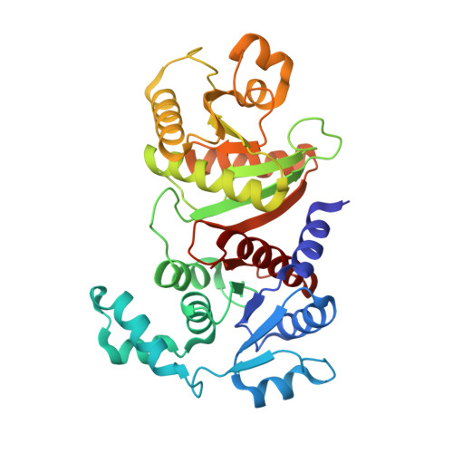
Crystal structure of phosphotransacetylase from the methanogenic archaeon Methanosarcina thermophila.

Iyer, P.P.Lawrence, S.H.Luther, K.B.Rajashankar, K.R.Yennawar, H.P.Ferry, J.G.Schindelin, H.

(2004) Structure 12: 559-567

  • DOI: https://doi.org/10.1016/j.str.2004.03.007
  • Primary Citation of Related Structures:  
    1QZT

  • PubMed Abstract: 

    Phosphotransacetylase (Pta) [EC 2.3.1.8] is ubiquitous in the carbon assimilation and energy-yielding pathways in anaerobic prokaryotes where it catalyzes the reversible transfer of the acetyl group from acetyl phosphate to CoA forming acetyl CoA and inorganic phosphate. The crystal structure of Pta from the methane-producing archaeon Methanosarcina thermophila, representing the first crystal structure of any Pta, was determined by multiwavelength anomalous diffraction at 2.7 A resolution. In solution and in the crystal, the enzyme forms a homodimer. Each monomer consists of two alpha/beta domains with a cleft along the domain boundary, which presumably contains the substrate binding sites. Comparison of the four monomers present in the asymmetric unit indicates substantial variations in the relative orientation of the two domains and the structure of the putative active site cleft. A search for structural homologs revealed the NADP(+)-dependent isocitrate and isopropylmalate dehydrogenases as the only homologs with a similar two-domain architecture.


  • Organizational Affiliation

    Department of Biochemistry and Molecular Biology, The Pennsylvania State University, University Park, PA 16802, USA.


Macromolecules
Find similar proteins by:  (by identity cutoff)  |  3D Structure
Entity ID: 1
MoleculeChains Sequence LengthOrganismDetailsImage
Phosphate acetyltransferase
A, B, C, D
333Methanosarcina thermophilaMutation(s): 0 
Gene Names: PTA
EC: 2.3.1.8
UniProt
Find proteins for P38503 (Methanosarcina thermophila)
Explore P38503 
Go to UniProtKB:  P38503
Entity Groups  
Sequence Clusters30% Identity50% Identity70% Identity90% Identity95% Identity100% Identity
UniProt GroupP38503
Sequence Annotations
Expand
  • Reference Sequence
Small Molecules
Ligands 1 Unique
IDChains Name / Formula / InChI Key2D Diagram3D Interactions
SO4
Query on SO4

Download Ideal Coordinates CCD File 
E [auth A]
F [auth A]
G [auth A]
H [auth A]
I [auth B]
E [auth A],
F [auth A],
G [auth A],
H [auth A],
I [auth B],
J [auth B],
K [auth B],
L [auth C],
M [auth C],
N [auth D],
O [auth D]
SULFATE ION
O4 S
QAOWNCQODCNURD-UHFFFAOYSA-L
Experimental Data & Validation

Experimental Data

  • Method: X-RAY DIFFRACTION
  • Resolution: 2.70 Å
  • R-Value Free: 0.286 
  • R-Value Work: 0.247 
  • R-Value Observed: 0.249 
  • Space Group: P 41
Unit Cell:
Length ( Å )Angle ( ˚ )
a = 115.345α = 90
b = 115.345β = 90
c = 129.366γ = 90
Software Package:
Software NamePurpose
REFMACrefinement
DENZOdata reduction
SCALEPACKdata scaling
MLPHAREphasing

Structure Validation

View Full Validation Report


Entry History 

Deposition Data

Revision History  (Full details and data files)

  • Version 1.0: 2004-06-22
    Type: Initial release
  • Version 1.1: 2008-04-29
    Changes: Version format compliance
  • Version 1.2: 2011-07-13
    Changes: Version format compliance
  • Version 1.3: 2018-06-27
    Changes: Data collection, Database references
  • Version 1.4: 2020-01-15
    Changes: Database references
  • Version 1.5: 2024-02-14
    Changes: Data collection, Database references, Derived calculations, Refinement description