Crystal structures of pinoresinol-lariciresinol and phenylcoumaran benzylic ether reductases and their relationship to isoflavone reductases.
Min, T., Kasahara, H., Bedgar, D.L., Youn, B., Lawrence, P.K., Gang, D.R., Halls, S.C., Park, H., Hilsenbeck, J.L., Davin, L.B., Lewis, N.G., Kang, C.(2003) J Biological Chem 278: 50714-50723
- PubMed: 13129921 Search on PubMed
- DOI: https://doi.org/10.1074/jbc.M308493200
- Primary Citation Related Structures:
1QYC, 1QYD - PubMed Abstract:
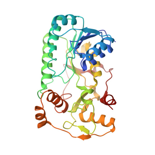
Despite the importance of plant lignans and isoflavonoids in human health protection (e.g. for both treatment and prevention of onset of various cancers) as well as in plant biology (e.g. in defense functions and in heartwood development), systematic studies on the enzymes involved in their biosynthesis have only recently begun. In this investigation, three NADPH-dependent aromatic alcohol reductases were comprehensively studied, namely pinoresinol-lariciresinol reductase (PLR), phenylcoumaran benzylic ether reductase (PCBER), and isoflavone reductase (IFR), which are involved in central steps to the various important bioactive lignans and isoflavonoids. Of particular interest was in determining how differing regio- and enantiospecificities are achieved with the different enzymes, despite each apparently going through similar enone intermediates. Initially, the three-dimensional x-ray crystal structures of both PLR_Tp1 and PCBER_Pt1 were solved and refined to 2.5 and 2.2 A resolutions, respectively. Not only do they share high gene sequence similarity, but their structures are similar, having a continuous alpha/beta NADPH-binding domain and a smaller substrate-binding domain. IFR (whose crystal structure is not yet obtained) was also compared (modeled) with PLR and PCBER and was deduced to have the same overall basic structure. The basis for the distinct enantio-specific and regio-specific reactions of PCBER, PLR, and IFR, as well as the reaction mechanism and participating residues involved (as identified by site-directed mutagenesis), are discussed.
- School of Molecular Biosciences, Washington State University, Pullman, Washington 99164-4660, USA.
Organizational Affiliation: