1QRV

CRYSTAL STRUCTURE OF THE COMPLEX OF HMG-D AND DNA


Experimental Data Snapshot

  • Method: X-RAY DIFFRACTION
  • Resolution: 2.20 Å
  • R-Value Free: 0.288 
  • R-Value Work: 0.238 
  • R-Value Observed: 0.238 

wwPDB Validation   3D Report Full Report


This is version 1.4 of the entry. See complete history


Literature

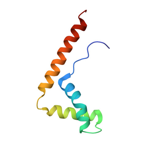
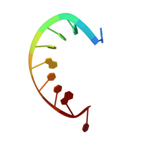
The structure of a chromosomal high mobility group protein-DNA complex reveals sequence-neutral mechanisms important for non-sequence-specific DNA recognition.

Murphy IV, F.V.Sweet, R.M.Churchill, M.E.

(1999) EMBO J 18: 6610-6618

  • DOI: https://doi.org/10.1093/emboj/18.23.6610
  • Primary Citation of Related Structures:  
    1QRV

  • PubMed Abstract: 

    The high mobility group (HMG) chromosomal proteins, which are common to all eukaryotes, bind DNA in a non-sequence-specific fashion to promote chromatin function and gene regulation. They interact directly with nucleosomes and are believed to be modulators of chromatin structure. They are also important in V(D)J recombination and in activating a number of regulators of gene expression, including p53, Hox transcription factors and steroid hormone receptors, by increasing their affinity for DNA. The X-ray crystal structure, at 2.2 A resolution, of the HMG domain of the Drosophila melanogaster protein, HMG-D, bound to DNA provides the first detailed view of a chromosomal HMG domain interacting with linear DNA and reveals the molecular basis of non-sequence-specific DNA recognition. Ser10 forms water-mediated hydrogen bonds to DNA bases, and Val32 with Thr33 partially intercalates the DNA. These two 'sequence-neutral' mechanisms of DNA binding substitute for base-specific hydrogen bonds made by equivalent residues of the sequence-specific HMG domain protein, lymphoid enhancer factor-1. The use of multiple intercalations and water-mediated DNA contacts may prove to be generally important mechanisms by which chromosomal proteins bind to DNA in the minor groove.


  • Organizational Affiliation

    Department of Pharmacology, The University of Colorado Health Sciences Center, 4200 E. Ninth Avenue, Denver, CO 80262, USA.


Macromolecules

Find similar proteins by:  (by identity cutoff)  |  3D Structure
Entity ID: 2
MoleculeChains Sequence LengthOrganismDetailsImage
HIGH MOBILITY GROUP PROTEIN DC [auth A],
D [auth B]
73Drosophila melanogasterMutation(s): 0 
UniProt
Find proteins for Q05783 (Drosophila melanogaster)
Explore Q05783 
Go to UniProtKB:  Q05783
Entity Groups  
Sequence Clusters30% Identity50% Identity70% Identity90% Identity95% Identity100% Identity
UniProt GroupQ05783
Sequence Annotations
Expand
  • Reference Sequence

Find similar nucleic acids by:  Sequence   |   3D Structure  

Entity ID: 1
MoleculeChains LengthOrganismImage
DNA (5'-D(*GP*CP*GP*AP*TP*AP*TP*CP*GP*C)-3')A [auth C],
B [auth D]
10N/A
Sequence Annotations
Expand
  • Reference Sequence
Small Molecules
Ligands 1 Unique
IDChains Name / Formula / InChI Key2D Diagram3D Interactions
NA
Query on NA

Download Ideal Coordinates CCD File 
E [auth C]SODIUM ION
Na
FKNQFGJONOIPTF-UHFFFAOYSA-N
Experimental Data & Validation

Experimental Data

  • Method: X-RAY DIFFRACTION
  • Resolution: 2.20 Å
  • R-Value Free: 0.288 
  • R-Value Work: 0.238 
  • R-Value Observed: 0.238 
  • Space Group: P 21 21 21
Unit Cell:
Length ( Å )Angle ( ˚ )
a = 43.74α = 90
b = 53.797β = 90
c = 86.843γ = 90
Software Package:
Software NamePurpose
SOLVEphasing
SHARPphasing
CNSrefinement
DENZOdata reduction
SCALEPACKdata scaling

Structure Validation

View Full Validation Report


Entry History 

Deposition Data

Revision History  (Full details and data files)

  • Version 1.0: 1999-12-18
    Type: Initial release
  • Version 1.1: 2008-04-27
    Changes: Version format compliance
  • Version 1.2: 2011-07-13
    Changes: Version format compliance
  • Version 1.3: 2018-01-31
    Changes: Database references
  • Version 1.4: 2024-02-14
    Changes: Data collection, Database references, Derived calculations