1QR4 | pdb_00001qr4

TWO FIBRONECTIN TYPE-III DOMAIN SEGMENT FROM CHICKEN TENASCIN


Experimental Data Snapshot

  • Method: X-RAY DIFFRACTION
  • Resolution: 2.55 Å
  • R-Value Free: 
    0.292 (Depositor) 
  • R-Value Work: 
    0.209 (Depositor), 0.220 (DCC) 
  • R-Value Observed: 
    0.209 (Depositor) 

Starting Model: experimental
View more details

wwPDB Validation 3D Report Full Report

Validation slider image for 1QR4

This is version 1.5 of the entry. See complete history

Literature

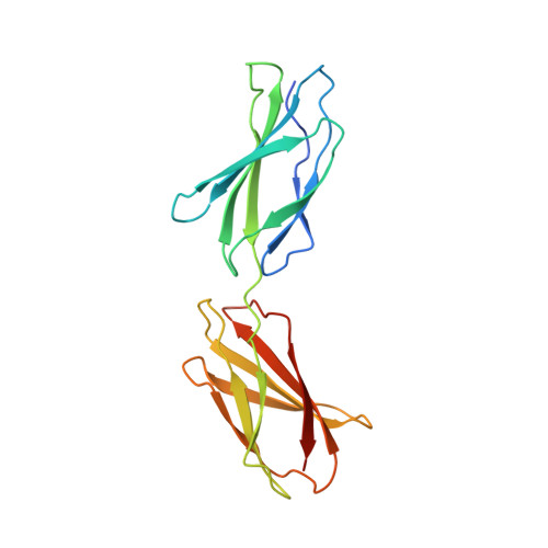
Purification, crystallization and preliminary crystallographic studies of a two fibronectin type-III domain segment from chicken tenascin encompassing the heparin- and contactin-binding regions.

Bisig, D.Weber, P.Vaughan, L.Winterhalter, K.H.Piontek, K.

(1999) Acta Crystallogr D Biol Crystallogr 55: 1069-1073

  • DOI: https://doi.org/10.1107/s090744499900284x
  • Primary Citation Related Structures: 
    1QR4

  • PubMed Abstract: 

    A fragment of chicken tenascin consisting of fibronectin type-III domains 5 and 6 has been expressed in Escherichia coli. After modifying a previously reported purification protocol, an electrophoretically homogeneous recombinant protein was obtained from which various crystal forms could be grown under identical conditions. Only one form was suitable for structure determination. These crystals belong to space group P21, with unit-cell parameters a = 45.2, b = 57.9, c = 72.2 A, beta = 91.4 degrees, and diffract to at least 2.6 A resolution using synchrotron radiation. From density measurements of the crystals, it was found that there are two molecules in the asymmetric unit. Diffraction data of native, two platinum-derivative and one palladium-derivative crystals were collected.


  • Organizational Affiliation
    • Laboratorium für Biochemie I, Swiss Federal Institute of Technology (ETH), CH-8092 Zürich, Switzerland.

Macromolecule Content 

  • Total Structure Weight: 40.52 kDa 
  • Atom Count: 2,787 
  • Modeled Residue Count: 350 
  • Deposited Residue Count: 372 
  • Unique protein chains: 1

Macromolecules

Find similar proteins by:|  3D Structure
Entity ID: 1
MoleculeChains  Sequence LengthOrganismDetailsImage
PROTEIN (TENASCIN)
A, B
186Gallus gallusMutation(s): 0 
UniProt
Find proteins for P10039 (Gallus gallus)
Explore P10039 
Go to UniProtKB:  P10039
Entity Groups
Sequence Clusters30% Identity50% Identity70% Identity90% Identity95% Identity100% Identity
UniProt GroupP10039
Sequence Annotations
Expand
Reference Sequence

Experimental Data & Validation

Experimental Data

  • Method: X-RAY DIFFRACTION
  • Resolution: 2.55 Å
  • R-Value Free:  0.292 (Depositor) 
  • R-Value Work:  0.209 (Depositor), 0.220 (DCC) 
  • R-Value Observed: 0.209 (Depositor) 
Space Group: P 1 21 1
Unit Cell:
Length ( Å )Angle ( ˚ )
a = 45.24α = 90
b = 57.94β = 91.4
c = 72.24γ = 90
Software Package:
Software NamePurpose
X-PLORmodel building
AMoREphasing
X-PLORrefinement
DENZOdata reduction
SCALEPACKdata scaling
X-PLORphasing

Structure Validation

View Full Validation Report


Entry History 

Deposition Data

Revision History  (Full details and data files)

  • Version 1.0: 1999-06-28
    Type: Initial release
  • Version 1.1: 2007-10-16
    Changes: Version format compliance
  • Version 1.2: 2011-07-13
    Changes: Version format compliance
  • Version 1.3: 2017-08-02
    Changes: Refinement description, Source and taxonomy
  • Version 1.4: 2019-12-25
    Changes: Advisory, Derived calculations
  • Version 1.5: 2023-08-16
    Changes: Data collection, Database references, Refinement description