1QR1

POOR BINDING OF A HER-2/NEU EPITOPE (GP2) TO HLA-A2.1 IS DUE TO A LACK OF INTERACTIONS IN THE CENTER OF THE PEPTIDE


Experimental Data Snapshot

  • Method: X-RAY DIFFRACTION
  • Resolution: 2.40 Å
  • R-Value Free: 0.284 
  • R-Value Work: 0.242 
  • R-Value Observed: 0.284 

wwPDB Validation   3D Report Full Report


This is version 1.6 of the entry. See complete history


Literature

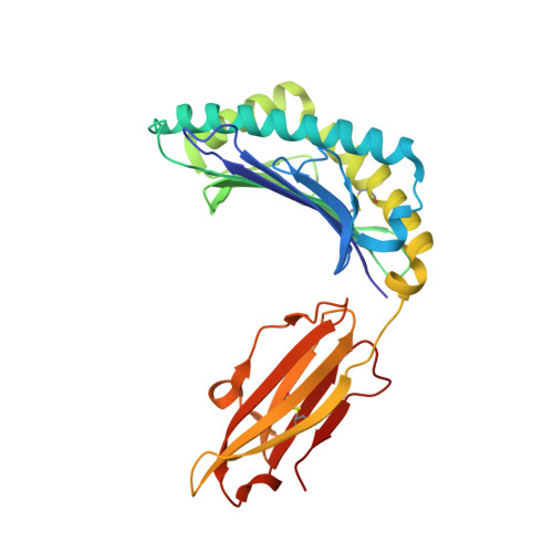
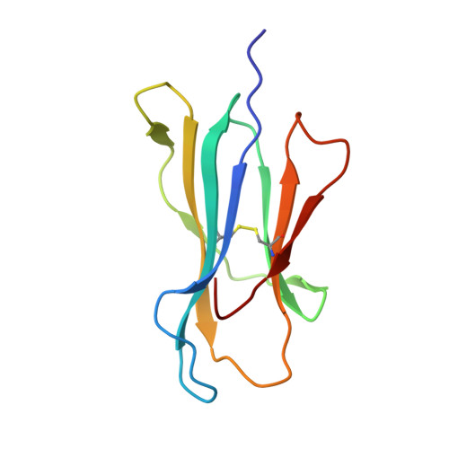
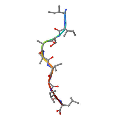
Poor binding of a HER-2/neu epitope (GP2) to HLA-A2.1 is due to a lack of interactions with the center of the peptide.

Kuhns, J.J.Batalia, M.A.Yan, S.Collins, E.J.

(1999) J Biol Chem 274: 36422-36427

  • DOI: https://doi.org/10.1074/jbc.274.51.36422
  • Primary Citation of Related Structures:  
    1QR1

  • PubMed Abstract: 

    Class I major histocompatibility complex (MHC) molecules bind short peptides derived from proteins synthesized within the cell. These complexes of peptide and class I MHC (pMHC) are transported from the endoplasmic reticulum to the cell surface. If a clonotypic T cell receptor expressed on a circulating T cell binds to the pMHC complex, the cell presenting the pMHC is killed. In this manner, some tumor cells expressing aberrant proteins are recognized and removed by the immune system. However, not all tumors are recognized efficiently. One reason hypothesized for poor T cell recognition of tumor-associated peptides is poor binding of those peptides to class I MHC molecules. Many peptides, derived from the proto-oncogene HER-2/neu have been shown to be recognized by cytotoxic T cells derived from HLA-A2(+) patients with breast cancer and other adenocarcinomas. Seven of these peptides were found to bind with intermediate to poor affinity. In particular, GP2 (HER-2/neu residues 654-662) binds very poorly even though it is predicted to bind well based upon the presence of the correct HLA-A2.1 peptide-binding motif. Altering the anchor residues to those most favored by HLA-A2.1 did not significantly improve binding affinity. The crystallographic structure shows that unlike other class I-peptide structures, the center of the peptide does not assume one specific conformation and does not make stabilizing contacts with the peptide-binding cleft.


  • Organizational Affiliation

    Department of Microbiology and Immunology, University of North Carolina, Chapel Hill, North Carolina 27599, USA.


Macromolecules
Find similar proteins by:  (by identity cutoff)  |  3D Structure
Entity ID: 1
MoleculeChains Sequence LengthOrganismDetailsImage
HLA-A2.1 HEAVY CHAIN
A, D
275Homo sapiensMutation(s): 0 
UniProt & NIH Common Fund Data Resources
Find proteins for P04439 (Homo sapiens)
Explore P04439 
Go to UniProtKB:  P04439
PHAROS:  P04439
GTEx:  ENSG00000206503 
Entity Groups  
Sequence Clusters30% Identity50% Identity70% Identity90% Identity95% Identity100% Identity
UniProt GroupP04439
Sequence Annotations
Expand
  • Reference Sequence
Find similar proteins by:  (by identity cutoff)  |  3D Structure
Entity ID: 2
MoleculeChains Sequence LengthOrganismDetailsImage
BETA-2 MICROGLOBULIN
B, E
100Homo sapiensMutation(s): 1 
UniProt & NIH Common Fund Data Resources
Find proteins for P61769 (Homo sapiens)
Explore P61769 
Go to UniProtKB:  P61769
PHAROS:  P61769
GTEx:  ENSG00000166710 
Entity Groups  
Sequence Clusters30% Identity50% Identity70% Identity90% Identity95% Identity100% Identity
UniProt GroupP61769
Sequence Annotations
Expand
  • Reference Sequence

Find similar proteins by:  Sequence   |   3D Structure  

Entity ID: 3
MoleculeChains Sequence LengthOrganismDetailsImage
GP2 PEPTIDE
C, F
9N/AMutation(s): 0 
UniProt & NIH Common Fund Data Resources
Find proteins for P04626 (Homo sapiens)
Explore P04626 
Go to UniProtKB:  P04626
PHAROS:  P04626
GTEx:  ENSG00000141736 
Entity Groups  
UniProt GroupP04626
Sequence Annotations
Expand
  • Reference Sequence
Experimental Data & Validation

Experimental Data

  • Method: X-RAY DIFFRACTION
  • Resolution: 2.40 Å
  • R-Value Free: 0.284 
  • R-Value Work: 0.242 
  • R-Value Observed: 0.284 
  • Space Group: P 1
Unit Cell:
Length ( Å )Angle ( ˚ )
a = 50.337α = 81.88
b = 63.609β = 76.25
c = 75.135γ = 77.83
Software Package:
Software NamePurpose
ADSCdata collection
DENZOdata reduction
SCALEPACKdata scaling
AMoREphasing
CNSrefinement
Adxvdata processing

Structure Validation

View Full Validation Report


Entry History 

Deposition Data

Revision History  (Full details and data files)

  • Version 1.0: 2000-01-01
    Type: Initial release
  • Version 1.1: 2008-04-27
    Changes: Version format compliance
  • Version 1.2: 2011-07-13
    Changes: Version format compliance
  • Version 1.3: 2017-02-08
    Changes: Structure summary
  • Version 1.4: 2017-10-04
    Changes: Advisory, Refinement description
  • Version 1.5: 2018-01-31
    Changes: Experimental preparation
  • Version 1.6: 2021-11-03
    Changes: Advisory, Database references