1QP1

KAPPA VARIABLE LIGHT CHAIN


Experimental Data Snapshot

  • Method: X-RAY DIFFRACTION
  • Resolution: 2.06 Å
  • R-Value Free: 0.212 
  • R-Value Work: 0.153 
  • R-Value Observed: 0.188 

wwPDB Validation   3D Report Full Report


This is version 1.3 of the entry. See complete history


Literature

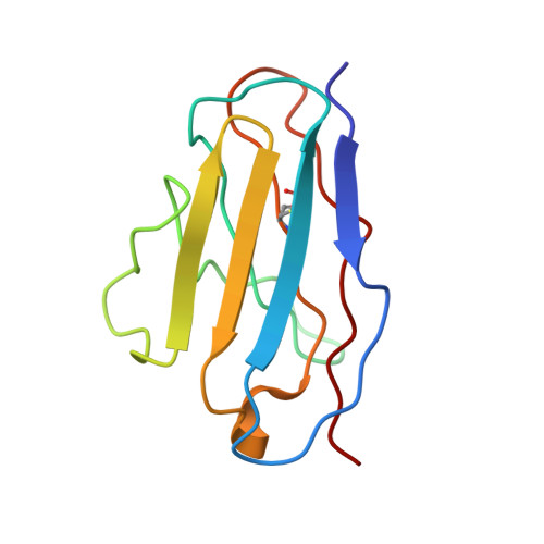
Molecular structure of the amyloid-forming protein kappa I Bre.

Steinrauf, L.K.Chiang, M.Y.Shiuan, D.

(1999) J Biochem 125: 422-429

  • DOI: 10.1093/oxfordjournals.jbchem.a022303
  • Primary Citation of Related Structures:  
    1QP1

  • PubMed Abstract: 
  • The molecular structure of the amyloid-forming Bence-Jones protein kappa I Bre has been determined by X-ray crystallography at 2.0 A resolution. The fragment from the kappa chain of immunoprotein contains 107 amino acid residues, and polymerizes in the crystal form into a giant helical spiral, surrounding a cylinder of water 50 A in diameter with a repeat of 77 ...

    The molecular structure of the amyloid-forming Bence-Jones protein kappa I Bre has been determined by X-ray crystallography at 2.0 A resolution. The fragment from the kappa chain of immunoprotein contains 107 amino acid residues, and polymerizes in the crystal form into a giant helical spiral, surrounding a cylinder of water 50 A in diameter with a repeat of 77.56 A, containing 12 kappa molecules, plus another 12 molecules from neighboring parallel spirals. The resulting structure has many features which have been found or suggested from studies on the protein fibrils found in amyloid deposits. From the results of the X-ray crystal structure a hypothesis is presented for the structure and formation of the amyloid fibril.


    Related Citations: 
    • Structure of an Immunoglubulin Kappa Variable Light Chain Associated with Primary Amyloidosis
      Steinrauf, L.K., Hamilton, J.A., Clawson, D., Liepnieks, J., Murrell, J., Benson, M.D.
      (1993) Am Cryst Assoc ,abstr Papers (annual Meeting) --: 128
    • Tertiary Structure of an Amyloid Immunoglobulin Light Chain Protein: A Proposed Model for Amyloid Fibril Formation
      Schormann, N., Murrell, J.R., Liepnieks, J.R., Benson, M.D.
      (1995) Proc Natl Acad Sci U S A 92: 9490

    Organizational Affiliation

    Department of Biochemistry and Molecular Biology, Indiana University School of Medicine, Indianapolis, Indiana, USA. lsteinra@www.ora.nsysu.edu.tw


Macromolecules
Find similar proteins by:  (by identity cutoff)  |  3D Structure
Entity ID: 1
MoleculeChainsSequence LengthOrganismDetailsImage
BENCE-JONES KAPPA I ANTIBODY BRE (LIGHT CHAIN)A, B, C107Homo sapiensMutation(s): 0 
Gene Names: IGKV1-33
UniProt
Find proteins for P01594 (Homo sapiens)
Explore P01594 
Go to UniProtKB:  P01594
Entity Groups  
Sequence Clusters30% Identity50% Identity70% Identity90% Identity95% Identity100% Identity
UniProt GroupP01594
Protein Feature View
Expand
  • Reference Sequence
Experimental Data & Validation

Experimental Data

  • Method: X-RAY DIFFRACTION
  • Resolution: 2.06 Å
  • R-Value Free: 0.212 
  • R-Value Work: 0.153 
  • R-Value Observed: 0.188 
  • Space Group: C 2 2 21
Unit Cell:
Length ( Å )Angle ( ˚ )
a = 143.99α = 90
b = 82.56β = 90
c = 77.56γ = 90
Software Package:
Software NamePurpose
X-PLORmodel building
SHELXL-97refinement
X-PLORphasing

Structure Validation

View Full Validation Report


Entry History 

Deposition Data

Revision History  (Full details and data files)

  • Version 1.0: 1999-06-17
    Type: Initial release
  • Version 1.1: 2008-04-27
    Changes: Version format compliance
  • Version 1.2: 2011-07-13
    Changes: Version format compliance
  • Version 1.3: 2017-10-04
    Changes: Refinement description