1QO3

Complex between NK cell receptor Ly49A and its MHC class I ligand H-2Dd


Experimental Data Snapshot

  • Method: X-RAY DIFFRACTION
  • Resolution: 2.30 Å
  • R-Value Free: 0.238 
  • R-Value Work: 0.198 
  • R-Value Observed: 0.198 

wwPDB Validation   3D Report Full Report


This is version 1.3 of the entry. See complete history


Literature

Crystal Structure of a Lectin-Like Natural Killer Cell Receptor Bound to its Mhc Class I Ligand

Tormo, J.Natarajan, K.Margulies, D.H.Mariuzza, R.A.

(1999) Nature 402: 623

  • DOI: https://doi.org/10.1038/45170
  • Primary Citation of Related Structures:  
    1QO3

  • PubMed Abstract: 

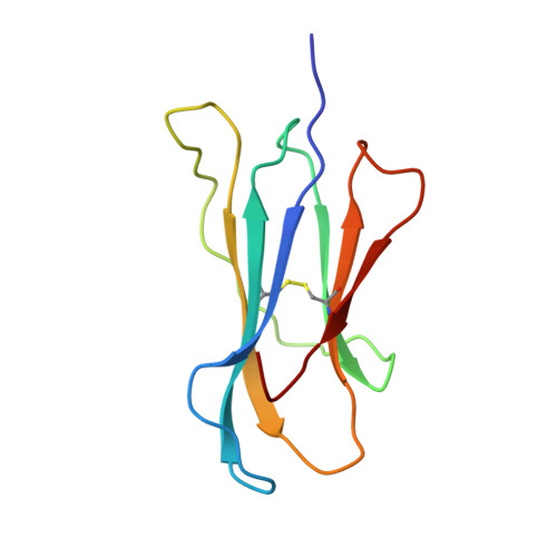
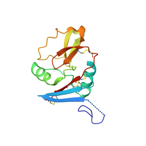
    Natural killer (NK) cell function is regulated by NK receptors that interact with MHC class I (MHC-I) molecules on target cells. The murine NK receptor Ly49A inhibits NK cell activity by interacting with H-2D(d) through its C-type-lectin-like NK receptor domain. Here we report the crystal structure of the complex between the Ly49A NK receptor domain and unglycosylated H-2D(d). The Ly49A dimer interacts extensively with two H-2D(d) molecules at distinct sites. At one interface, a single Ly49A subunit contacts one side of the MHC-I peptide-binding platform, presenting an open cavity towards the conserved glycosylation site on the H-2D(d) alpha2 domain. At a second, larger interface, the Ly49A dimer binds in a region overlapping the CD8-binding site. The smaller interface probably represents the interaction between Ly49A on the NK cell and MHC-I on the target cell, whereas the larger one suggests an interaction between Ly49A and MHC-I on the NK cell itself. Both Ly49A binding sites on MHC-I are spatially distinct from that of the T-cell receptor.


  • Organizational Affiliation

    Center for Advanced Research in Biotechnology, University of Maryland Biotechnology Institute, Rockville 20850, USA.


Macromolecules
Find similar proteins by:  (by identity cutoff)  |  3D Structure
Entity ID: 1
MoleculeChains Sequence LengthOrganismDetailsImage
MHC CLASS I H-2DD HEAVY CHAIN277Mus musculusMutation(s): 0 
Gene Names: H-2D
UniProt
Find proteins for P01900 (Mus musculus)
Explore P01900 
Go to UniProtKB:  P01900
Entity Groups  
Sequence Clusters30% Identity50% Identity70% Identity90% Identity95% Identity100% Identity
UniProt GroupP01900
Sequence Annotations
Expand
  • Reference Sequence
Find similar proteins by:  (by identity cutoff)  |  3D Structure
Entity ID: 2
MoleculeChains Sequence LengthOrganismDetailsImage
BETA-2-MICROGLOBULIN100Mus musculusMutation(s): 1 
UniProt & NIH Common Fund Data Resources
Find proteins for P01887 (Mus musculus)
Explore P01887 
Go to UniProtKB:  P01887
IMPC:  MGI:88127
Entity Groups  
Sequence Clusters30% Identity50% Identity70% Identity90% Identity95% Identity100% Identity
UniProt GroupP01887
Sequence Annotations
Expand
  • Reference Sequence
Find similar proteins by:  (by identity cutoff)  |  3D Structure
Entity ID: 3
MoleculeChains Sequence LengthOrganismDetailsImage
LY49A
C, D
137Mus musculusMutation(s): 3 
UniProt
Find proteins for P20937 (Mus musculus)
Explore P20937 
Go to UniProtKB:  P20937
Entity Groups  
Sequence Clusters30% Identity50% Identity70% Identity90% Identity95% Identity100% Identity
UniProt GroupP20937
Sequence Annotations
Expand
  • Reference Sequence

Find similar proteins by:  Sequence   |   3D Structure  

Entity ID: 4
MoleculeChains Sequence LengthOrganismDetailsImage
HIV ENVELOPE GLYCOPROTEIN 120 PEPTIDEE [auth P]10Human immunodeficiency virusMutation(s): 0 
UniProt
Find proteins for P04582 (Human immunodeficiency virus type 1 group M subtype B (isolate BH8))
Explore P04582 
Go to UniProtKB:  P04582
Entity Groups  
Sequence Clusters30% Identity50% Identity70% Identity90% Identity95% Identity100% Identity
UniProt GroupP04582
Sequence Annotations
Expand
  • Reference Sequence
Small Molecules
Experimental Data & Validation

Experimental Data

  • Method: X-RAY DIFFRACTION
  • Resolution: 2.30 Å
  • R-Value Free: 0.238 
  • R-Value Work: 0.198 
  • R-Value Observed: 0.198 
  • Space Group: P 21 21 21
Unit Cell:
Length ( Å )Angle ( ˚ )
a = 78.963α = 90
b = 96.957β = 90
c = 99.318γ = 90
Software Package:
Software NamePurpose
CNSrefinement
DENZOdata reduction
SCALEPACKdata scaling
CNSphasing

Structure Validation

View Full Validation Report


Entry History 

Deposition Data

Revision History  (Full details and data files)

  • Version 1.0: 2000-01-02
    Type: Initial release
  • Version 1.1: 2013-01-23
    Changes: Data collection, Database references, Derived calculations, Non-polymer description, Other, Refinement description, Structure summary, Version format compliance
  • Version 1.2: 2015-04-15
    Changes: Database references
  • Version 1.3: 2023-12-13
    Changes: Data collection, Database references, Derived calculations, Other, Refinement description