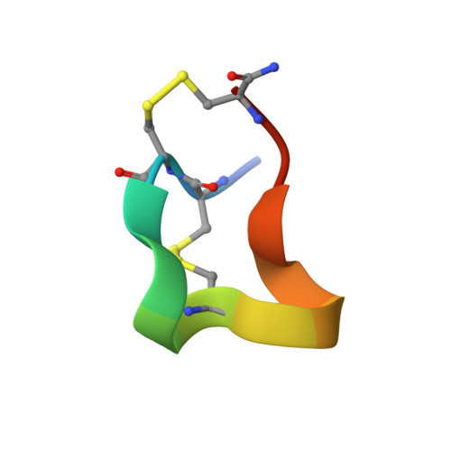
1QMW

Solution structure of alpha-conotoxin SI


Experimental Data Snapshot

  • Method: SOLUTION NMR
  • Conformers Calculated: 50 
  • Conformers Submitted: 36 
  • Selection Criteria: LOWEST ENERGY STRUCTURES 

wwPDB Validation   3D Report Full Report


This is version 1.4 of the entry. See complete history


Literature

Solution Structure of Alpha-Conotoxin Si

Benie, A.J.Whitford, D.Hargittai, B.Barany, G.Janes, R.W.

(2000) FEBS Lett 476: 287

  • DOI: https://doi.org/10.1016/s0014-5793(00)01724-5
  • Primary Citation of Related Structures:  
    1QMW

  • PubMed Abstract: 
  • The nuclear magnetic resonance solution structure of alpha-conotoxin SI has been determined at pH 4.2. The 36 lowest energy structures show that alpha-conotoxin SI exists in a single major solution conformation and is stabilized by six hydrogen bonds. Comparisons are made between the SI solution structure and the solution and crystal structures of alpha-conotoxin GI ...

    The nuclear magnetic resonance solution structure of alpha-conotoxin SI has been determined at pH 4.2. The 36 lowest energy structures show that alpha-conotoxin SI exists in a single major solution conformation and is stabilized by six hydrogen bonds. Comparisons are made between the SI solution structure and the solution and crystal structures of alpha-conotoxin GI. Surprisingly, a high degree of similarity between the backbone conformations of the GI crystal and the SI solution structures is seen in the region of lowest sequence homology, namely residues Gly-8 to Ser-12. This similarity is more surprising when considering that in SI a proline replaces the Arg-9 found in GI. The correspondence in conformation in this region provides the definitive evidence that it is the loss of the arginine basic charge at residue 9 which determines the differences in toxicity between GI and SI, rather than any changes in conformation induced by the cyclic proline residue.


    Organizational Affiliation

    Molecular and Cellular Biology, St. Bartholomew's and the Royal London School of Medicine and Dentistry, University of London, UK.


Macromolecules

Find similar proteins by:  Sequence   |   3D Structure  

Entity ID: 1
MoleculeChainsSequence LengthOrganismDetailsImage
ALPHA-CONOTOXIN SI14Conus striatusMutation(s): 0 
Membrane Entity: Yes 
UniProt
Find proteins for P15471 (Conus striatus)
Explore P15471 
Go to UniProtKB:  P15471
Entity Groups  
Sequence Clusters30% Identity50% Identity70% Identity90% Identity95% Identity100% Identity
UniProt GroupP15471
Protein Feature View
Expand
  • Reference Sequence
Experimental Data & Validation

Experimental Data

  • Method: SOLUTION NMR
  • Conformers Calculated: 50 
  • Conformers Submitted: 36 
  • Selection Criteria: LOWEST ENERGY STRUCTURES 

Structure Validation

View Full Validation Report


Entry History 

Deposition Data

Revision History  (Full details and data files)

  • Version 1.0: 2000-08-25
    Type: Initial release
  • Version 1.1: 2011-05-08
    Changes: Version format compliance
  • Version 1.2: 2011-07-13
    Changes: Version format compliance
  • Version 1.3: 2020-01-15
    Changes: Derived calculations, Other
  • Version 1.4: 2023-06-14
    Changes: Database references, Other