1QLM | pdb_00001qlm

The crystal structure of methenyltetrahydromethanopterin cyclohydrolase from the hyperthermophilic archaeon Methanopyrus kandleri


Experimental Data Snapshot

  • Method: X-RAY DIFFRACTION
  • Resolution: 2.00 Å
  • R-Value Free: 
    0.219 (Depositor), 0.225 (DCC) 
  • R-Value Work: 
    0.198 (Depositor), 0.206 (DCC) 
  • R-Value Observed: 
    0.198 (Depositor) 

wwPDB Validation 3D Report Full Report

Validation slider image for 1QLM

This is version 1.3 of the entry. See complete history

Literature

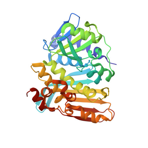
The Crystal Structure of Methenyltetrahydromethano- Pterin Cyclohydrolase from the Hyperthermophilic Archaeon Methanopyrus Kandleri

Grabarse, W.Vaupel, M.Vorholt, J.A.Shima, S.Thauer, R.K.Wittershagen, A.Bourenkov, G.Bartunik, H.D.Ermler, U.

(1999) Structure 7: 1257

  • DOI: https://doi.org/10.1016/s0969-2126(00)80059-3
  • Primary Citation Related Structures: 
    1QLM

  • PubMed Abstract: 

    The reduction of carbon dioxide to methane in methanogenic archaea involves the tetrahydrofolate analogue tetrahydromethanopterin (H(4)MPT) as a C(1) unit carrier. In the third step of this reaction sequence, N(5)-formyl-H(4)MPT is converted to methenyl-H(4)MPT(+) by the enzyme methenyltetrahydromethanopterin cyclohydrolase. The cyclohydrolase from the hyperthermophilic archaeon Methanopyrus kandleri (Mch) is extremely thermostable and adapted to a high intracellular concentration of lyotropic salts. Mch was crystallized and its structure solved at 2.0 A resolution using a combination of the single isomorphous replacement (SIR) and multiple anomalous dispersion (MAD) techniques. The structure of the homotrimeric enzyme reveals a new alpha/beta fold that is composed of two domains forming a large sequence-conserved pocket between them. Two phosphate ions were found in and adjacent to this pocket, respectively; the latter is displaced by the phosphate moiety of the substrate formyl-H(4)MPT according to a hypothetical model of the substrate binding. Although the exact position of the substrate is not yet known, the residues lining the active site of Mch could be tentatively assigned. Comparison of Mch with the tetrahydrofolate-specific cyclohydrolase/dehydrogenase reveals similarities in domain arrangement and in some active-site residues, whereas the fold appears to be different. The adaptation of Mch to high salt concentrations and high temperatures is reflected by the excess of acidic residues at the trimer surface and by the higher oligomerization state of Mch compared with its mesophtic counterparts.


  • Organizational Affiliation
    • Max-Planck-Institut für terrestrische Mikrobiologie, Karl-von-Frisch-Strasse, 35043, Marburg, Max-Planck-Institut für Biophysik, Heinrich-Hoffmann-Strasse 7, 60528, Frankurt, Germany.

Macromolecule Content 

  • Total Structure Weight: 34.38 kDa 
  • Atom Count: 2,642 
  • Modeled Residue Count: 316 
  • Deposited Residue Count: 316 
  • Unique protein chains: 1

Macromolecules

Find similar proteins by:|  3D Structure
Entity ID: 1
MoleculeChains  Sequence LengthOrganismDetailsImage
METHENYLTETRAHYDROMETHANOPTERIN CYCLOHYDROLASE316Methanopyrus kandleriMutation(s): 0 
EC: 3.5.4.27
UniProt
Find proteins for P94954 (Methanopyrus kandleri (strain AV19 / DSM 6324 / JCM 9639 / NBRC 100938))
Explore P94954 
Go to UniProtKB:  P94954
Entity Groups
Sequence Clusters30% Identity50% Identity70% Identity90% Identity95% Identity100% Identity
UniProt GroupP94954
Sequence Annotations
Expand
Reference Sequence

Experimental Data & Validation

Experimental Data

  • Method: X-RAY DIFFRACTION
  • Resolution: 2.00 Å
  • R-Value Free:  0.219 (Depositor), 0.225 (DCC) 
  • R-Value Work:  0.198 (Depositor), 0.206 (DCC) 
  • R-Value Observed: 0.198 (Depositor) 
Space Group: P 63 2 2
Unit Cell:
Length ( Å )Angle ( ˚ )
a = 125.9α = 90
b = 125.9β = 90
c = 172.4γ = 120
Software Package:
Software NamePurpose
CNSrefinement
DENZOdata reduction
SCALEPACKdata scaling
CNSphasing

Structure Validation

View Full Validation Report


Entry History 

Deposition Data

  • Released Date: 1999-09-20 
  • Deposition Author(s): Grabarse, W.

Revision History  (Full details and data files)

  • Version 1.0: 1999-09-20
    Type: Initial release
  • Version 1.1: 2012-11-14
    Changes: Database references, Derived calculations, Non-polymer description, Other, Refinement description, Structure summary, Version format compliance
  • Version 1.2: 2017-07-05
    Changes: Data collection
  • Version 1.3: 2024-05-08
    Changes: Advisory, Data collection, Database references, Derived calculations, Other