The Molecular Structure and Structural Transition of the Alpha-Helical Capsid in Filamentous Bacteriophage Pf1
Welsh, L.C., Symmons, M.F., Marvin, D.A.(2000) Acta Crystallogr D Biol Crystallogr 56: 137
- PubMed: 10666593 Search on PubMed
- DOI: https://doi.org/10.1107/s0907444999015334
- Primary Citation Related Structures:
1QL1, 1QL2 - PubMed Abstract:
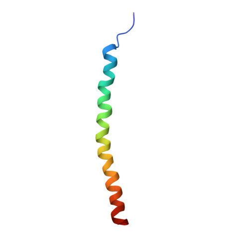
The major coat protein in the capsid of Pf1 filamentous bacteriophage (Inovirus) forms a helical assembly of about 7000 identical protein subunits, each of which contains 46 amino-acid residues and can be closely approximated by a single gently curved alpha-helix. Since the viral DNA occupies the core of the tubular capsid and appears to make no significant specific interactions with the capsid proteins, the capsid is a simple model system for the study of the static and dynamic properties of alpha-helix assembly. The capsid undergoes a reversible temperature-induced structural transition at about 283 K between two slightly different helix forms. The two forms can coexist without an intermediate state, consistent with a first-order structural phase transition. The molecular model of the higher temperature form was refined using improved X-ray fibre diffraction data and new refinement and validation methods. The refinement indicates that the two forms are related by a change in the orientation of the capsid subunits within the virion, without a significant change in local conformation of the subunits. On the higher temperature diffraction pattern there is a region of observed intensity that is not consistent with a simple helix of identical subunits; it is proposed that the structure involves groups of three subunits which each have a slightly different orientation within the group. The grouping of subunits suggests that a change in subunit libration frequency could be the basis of the Pf1 structural transition; calculations from the model are used to explore this idea.
- Cambridge Centre for Molecular Recognition, Department of Biochemistry, University of Cambridge, 80 Tennis Court Road, Cambridge CB2 1GA, England.
Organizational Affiliation: