1QL0 | pdb_00001ql0

Sm Endonuclease from Seratia marcenscens at atomic resolution


Experimental Data Snapshot

  • Method: X-RAY DIFFRACTION
  • Resolution: 1.10 Å
  • R-Value Free: 
    0.147 (Depositor) 
  • R-Value Work: 
    0.139 (DCC) 
  • R-Value Observed: 
    0.128 (Depositor) 

wwPDB Validation 3D Report Full Report

Validation slider image for 1QL0

This is version 1.3 of the entry. See complete history

Literature

Atomic Structure of the Serratia Marcescens Endonuclease at 1.1 A Resolution and the Enzyme Reaction Mechanism.

Shlyapnikov, S.V.Lunin, V.V.Perbandt, M.Polyakov, K.M.Lunin, V.Y.Levdikov, V.M.Betzel, C.Mikhailov, A.M.

(2000) Acta Crystallogr D Biol Crystallogr 56: 567

  • DOI: https://doi.org/10.1107/s090744490000322x
  • Primary Citation Related Structures: 
    1G8T, 1QL0

  • PubMed Abstract: 

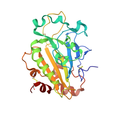
    The three-dimensional crystal structure of Serratia marcescens endonuclease has been refined at 1.1 A resolution to an R factor of 12.9% and an R(free) of 15.6% with the use of anisotropic temperature factors. The model contains 3694 non-H atoms, 715 water molecules, four sulfate ions and two Mg(2+)-binding sites at the active sites of the homodimeric protein. It is shown that the magnesium ion linked to the active-site Asn119 of each monomer is surrounded by five water molecules and shows an octahedral coordination geometry. The temperature factors for the bound Mg(2+) ions in the A and B subunits are 7.08 and 4.60 A(2), respectively, and the average temperature factors for the surrounding water molecules are 12.13 and 10.3 A(2), respectively. In comparison with earlier structures, alternative side-chain conformations are defined for 51 residues of the dimer, including the essential active-site residue Arg57. A plausible mechanism of enzyme function is proposed based on the high-resolution S. marcescens nuclease structure, the functional characteristics of the natural and mutational forms of the enzyme and consideration of its structural analogy with homing endo-nuclease I-PpoI.


  • Organizational Affiliation
    • Engelhardt Institute of Molecular Biology, Russian Academy of Sciences, Vavilov Str. 32, Moscow 117984, Russia.

Macromolecule Content 

  • Total Structure Weight: 52.64 kDa 
  • Atom Count: 4,541 
  • Modeled Residue Count: 482 
  • Deposited Residue Count: 482 
  • Unique protein chains: 1

Macromolecules

Find similar proteins by:|  3D Structure
Entity ID: 1
MoleculeChains  Sequence LengthOrganismDetailsImage
NUCLEASE
A, B
241Serratia marcescensMutation(s): 0 
EC: 3.1.30.2
UniProt
Find proteins for P13717 (Serratia marcescens)
Explore P13717 
Go to UniProtKB:  P13717
Entity Groups
Sequence Clusters30% Identity50% Identity70% Identity90% Identity95% Identity100% Identity
UniProt GroupP13717
Sequence Annotations
Expand
Reference Sequence

Experimental Data & Validation

Experimental Data

  • Method: X-RAY DIFFRACTION
  • Resolution: 1.10 Å
  • R-Value Free:  0.147 (Depositor) 
  • R-Value Work:  0.139 (DCC) 
  • R-Value Observed: 0.128 (Depositor) 
Space Group: P 21 21 2
Unit Cell:
Length ( Å )Angle ( ˚ )
a = 106.39α = 90
b = 73.67β = 90
c = 68.12γ = 90
Software Package:
Software NamePurpose
SHELXL-97refinement
SHELXL-97phasing

Structure Validation

View Full Validation Report


Entry History 

Deposition Data

Revision History  (Full details and data files)

  • Version 1.0: 2000-05-07
    Type: Initial release
  • Version 1.1: 2014-03-12
    Changes: Database references, Derived calculations, Non-polymer description, Other, Source and taxonomy, Structure summary, Version format compliance
  • Version 1.2: 2019-07-24
    Changes: Data collection
  • Version 1.3: 2024-10-16
    Changes: Data collection, Database references, Derived calculations, Other, Structure summary