1QKR

Crystal structure of the vinculin tail and a pathway for activation


Experimental Data Snapshot

  • Method: X-RAY DIFFRACTION
  • Resolution: 1.80 Å
  • R-Value Free: 0.248 
  • R-Value Work: 0.200 
  • R-Value Observed: 0.200 

wwPDB Validation   3D Report Full Report


This is version 1.2 of the entry. See complete history


Literature

Crystal Structure of the Vinculin Tail and a Pathway for Activation

Bakolitsa, C.De Pereda, J.M.Bagshaw, C.R.Critchley, D.R.Liddington, R.C.

(1999) Cell 99: 603

  • DOI: https://doi.org/10.1016/s0092-8674(00)81549-4
  • Primary Citation of Related Structures:  
    1QKR

  • PubMed Abstract: 

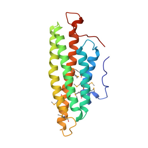
    Vinculin plays a dynamic role in the assembly of the actin cytoskeleton. A strong interaction between its head and tail domains that regulates binding to other cytoskeletal components is disrupted by acidic phospholipids. Here, we present the crystal structure of the vinculin tail, residues 879-1066. Five amphipathic helices form an antiparallel bundle that resembles exchangeable apolipoproteins. A C-terminal arm wraps across the base of the bundle and emerges as a hydrophobic hairpin surrounded by a collar of basic residues, adjacent to the N terminus. We show that the C-terminal arm is required for binding to acidic phospholipids but not to actin, and that binding either ligand induces conformational changes that may represent the first step in activation.


  • Organizational Affiliation

    Department of Biochemistry, University of Leicester, United Kingdom.


Macromolecules
Find similar proteins by:  (by identity cutoff)  |  3D Structure
Entity ID: 1
MoleculeChains Sequence LengthOrganismDetailsImage
VINCULIN
A, B
188Gallus gallusMutation(s): 9 
Gene Names: VCL
UniProt
Find proteins for P12003 (Gallus gallus)
Explore P12003 
Go to UniProtKB:  P12003
Entity Groups  
Sequence Clusters30% Identity50% Identity70% Identity90% Identity95% Identity100% Identity
UniProt GroupP12003
Sequence Annotations
Expand
  • Reference Sequence
Small Molecules
Modified Residues  1 Unique
IDChains TypeFormula2D DiagramParent
MSE
Query on MSE
A, B
L-PEPTIDE LINKINGC5 H11 N O2 SeMET
Experimental Data & Validation

Experimental Data

  • Method: X-RAY DIFFRACTION
  • Resolution: 1.80 Å
  • R-Value Free: 0.248 
  • R-Value Work: 0.200 
  • R-Value Observed: 0.200 
  • Space Group: P 1 21 1
Unit Cell:
Length ( Å )Angle ( ˚ )
a = 39.6α = 90
b = 87.77β = 91.27
c = 50.63γ = 90
Software Package:
Software NamePurpose
DENZOdata reduction
SCALEPACKdata scaling
CNSphasing
SOLVEphasing
CNSrefinement

Structure Validation

View Full Validation Report


Entry History 

Deposition Data

Revision History  (Full details and data files)

  • Version 1.0: 2000-08-04
    Type: Initial release
  • Version 1.1: 2011-05-08
    Changes: Version format compliance
  • Version 1.2: 2011-07-13
    Changes: Version format compliance