1QFC

STRUCTURE OF RAT PURPLE ACID PHOSPHATASE


Experimental Data Snapshot

  • Method: X-RAY DIFFRACTION
  • Resolution: 2.70 Å
  • R-Value Free: 0.281 
  • R-Value Work: 0.234 

wwPDB Validation   3D Report Full Report


This is version 1.3 of the entry. See complete history


Literature

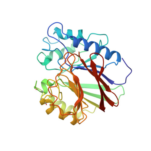
Crystal structure of a mammalian purple acid phosphatase.

Uppenberg, J.Lindqvist, F.Svensson, C.Ek-Rylander, B.Andersson, G.

(1999) J Mol Biol 290: 201-211

  • DOI: 10.1006/jmbi.1999.2896
  • Primary Citation of Related Structures:  
    1QFC

  • PubMed Abstract: 
  • Tartrate-resistant acid phosphatase (TRAP) is a mammalian di-iron- containing enzyme that belongs to the family of purple acid phosphatases (PAP). It is highly expressed in a limited number of tissues, predominantly in bone-resorbing osteoclasts and in macrophages of spleen ...

    Tartrate-resistant acid phosphatase (TRAP) is a mammalian di-iron- containing enzyme that belongs to the family of purple acid phosphatases (PAP). It is highly expressed in a limited number of tissues, predominantly in bone-resorbing osteoclasts and in macrophages of spleen. We have determined the crystal structure of rat TRAP in complex with a phosphate ion to 2.7 A resolution. The fold resembles that of the catalytic domain of kidney bean purple acid phosphatase (KBPAP), although the sequence similarity is limited to the active site residues. A surface loop near the active site is absent due to proteolysis, leaving the active-site easily accessible from the surrounding solvent. This, we believe, gives a structural explanation for the observed proteolytic activation of TRAP. The current structure was determined at a relatively high pH and without any external reducing agents. It is likely that it represents an oxidized and therefore catalytically inactive form of the enzyme.


    Organizational Affiliation

    Department of Structural Chemistry, Pharmacia and Upjohn, Lindhagensgatan 133, Stockholm, S-112 87, Sweden. jonas.uppenberg@eu.pnu.com


Macromolecules
Find similar proteins by:  (by identity cutoff)  |  3D Structure
Entity ID: 1
MoleculeChainsSequence LengthOrganismDetailsImage
PROTEIN (PURPLE ACID PHOSPHATASE)A306Rattus norvegicusMutation(s): 0 
Gene Names: Acp5
EC: 3.1.3.2
UniProt
Find proteins for P29288 (Rattus norvegicus)
Explore P29288 
Go to UniProtKB:  P29288
Entity Groups  
Sequence Clusters30% Identity50% Identity70% Identity90% Identity95% Identity100% Identity
UniProt GroupP29288
Protein Feature View
Expand
  • Reference Sequence
Small Molecules
Ligands 3 Unique
IDChainsName / Formula / InChI Key2D Diagram3D Interactions
NAG
Query on NAG

Download Ideal Coordinates CCD File 
B [auth A]2-acetamido-2-deoxy-beta-D-glucopyranose
C8 H15 N O6
OVRNDRQMDRJTHS-FMDGEEDCSA-N
 Ligand Interaction
PO4
Query on PO4

Download Ideal Coordinates CCD File 
E [auth A]PHOSPHATE ION
O4 P
NBIIXXVUZAFLBC-UHFFFAOYSA-K
 Ligand Interaction
FE
Query on FE

Download Ideal Coordinates CCD File 
C [auth A],
D [auth A]
FE (III) ION
Fe
VTLYFUHAOXGGBS-UHFFFAOYSA-N
 Ligand Interaction
Experimental Data & Validation

Experimental Data

  • Method: X-RAY DIFFRACTION
  • Resolution: 2.70 Å
  • R-Value Free: 0.281 
  • R-Value Work: 0.234 
  • Space Group: I 41
Unit Cell:
Length ( Å )Angle ( ˚ )
a = 116.38α = 90
b = 116.38β = 90
c = 63.27γ = 90
Software Package:
Software NamePurpose
DENZOdata reduction
SCALEPACKdata scaling
MLPHAREphasing
SHARPphasing
CNSrefinement

Structure Validation

View Full Validation Report


Entry History 

Deposition Data

Revision History  (Full details and data files)

  • Version 1.0: 2000-04-10
    Type: Initial release
  • Version 1.1: 2008-04-26
    Changes: Version format compliance
  • Version 1.2: 2011-07-13
    Changes: Non-polymer description, Version format compliance
  • Version 1.3: 2020-07-29
    Type: Remediation
    Reason: Carbohydrate remediation
    Changes: Data collection, Derived calculations, Structure summary