Crystal structure of epidoxorubicin-formaldehyde virtual crosslink of DNA and evidence for its formation in human breast-cancer cells.
Podell, E.R., Harrington, D.J., Taatjes, D.J., Koch, T.H.(1999) Acta Crystallogr D Biol Crystallogr 55: 1516-1523
- PubMed: 10489446 Search on PubMed
- DOI: https://doi.org/10.1107/s0907444999008161
- Primary Citation Related Structures:
1QDA - PubMed Abstract:
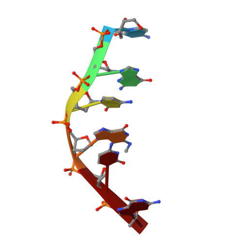
Epidoxorubicin and daunorubicin are proposed to be cytotoxic to tumor cells by catalyzing production of formaldehyde through redox cycling and using the formaldehyde for covalent attachment to DNA at G bases. The crystal structure of epidoxorubicin covalently bound to a d(CGCGCG) oligomer was determined to 1.6 A resolution. The structure reveals slightly distorted B-form DNA bearing two molecules of epidoxorubicin symmetrically intercalated at the termini, with each covalently attached from its N3' to N2 of a G base via a CH2 group from the formaldehyde. The structure is analogous to daunorubicin covalently bound to d(CGCGCG) determined previously, except for additional hydrogen bonding from the epimeric O4' to O2 of a C base. The role of drug-DNA covalent bonding in cells was investigated with synthetic epidoxorubicin-formaldehyde conjugate (Epidoxoform) and synthetic daunorubicin-formaldehyde conjugate (Daunoform). Uptake and location of drug fluorophore in doxorubicin-resistant human breast-cancer cells (MCF-7/ADR cells) was observed by fluorescence microscopy and flow cytometry. The fluorophore of Daunoform appeared more rapidly in cells and was released more rapidly from cells than the fluorophore of Epidoxoform over a 3 h exposure period. The fluorophore appeared predominantly in the nucleus of cells treated with both conjugates. The difference in uptake is explained in terms of the slower rate of hydrolysis of Epidoxoform to the species reactive with DNA and a proposed slower release from DNA based upon the crystal structures.
- Department of Chemistry and Biochemistry, University of Colorado, Boulder, Colorado 80309-0215, USA.
Organizational Affiliation: