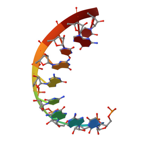
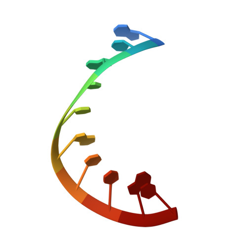
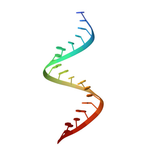
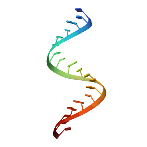
Crystal structures of two plasmid copy control related RNA duplexes: An 18 base pair duplex at 1.20 A resolution and a 19 base pair duplex at 1.55 A resolution.
Klosterman, P.S., Shah, S.A., Steitz, T.A.(1999) Biochemistry 38: 14784-14792
- PubMed: 10555960 Search on PubMed
- DOI: https://doi.org/10.1021/bi9912793
- Primary Citation Related Structures:
1QC0, 1QCU - PubMed Abstract:
The structures of two RNA duplexes, whose sequences correspond to portions of the ColE1 plasmid copy control RNA I and RNA II, have been determined. Crystals containing the 18mers 5'-CA CCGUUGGUAGCGGUGC-3' and 5'-CACCGCUACCAACGGUGC-3' diffract to 1.20 A resolution while those containing the 19mers 5'-GCACCGUUGGUAGCGGUGC-3' and 5'-GCACCGCUACCAACGGUGC-3' diffract to 1.55 A resolution. Both duplexes are standard A form, with Watson-Crick base pairing throughout. Use of anisotropic atomic displacement factors in refinement of the 1.20 A structure dramatically improved refinement statistics, resulting in a final R(free) of 15.0% and a crystallographic R-factor of 11.6%. Perhaps surprisingly, these crystals of the 18 base pair RNA exhibit a 36-fold static disorder, resulting in a structure with a single sugar-phosphate backbone conformation and an averaged base composition at each residue. Since the sugar-phosphate backbone structure is identical in the 36 different nucleotides that are superimposed, there can be no sequence-dependent variation in the structure. The average ribose pucker amplitude is 45.8 degrees for the 18 base pair structure and 46.4 degrees for the 19 base pair structure; these values are respectively 19% and 20% larger than the average pucker amplitude reported from nucleoside crystal structures. A standard RNA water structure, based on analysis of the hydration of these crystal structures and that of the TAR RNA stem [Ippolito, J. A., and Steitz, T. A. (1998) Proc. Natl. Acad. Sci. U.S.A. 95, 9819-9824], has been derived, which has allowed us to predict water positions in lower resolution RNA crystal structures. We report a new RNA packing motif, in which three pro-S(p) phosphate oxygens interact with an ammonium ion.
- Department of Molecular Biophysics and Biochemistry, Yale University, 266 Whitney Avenue, New Haven, Connecticut 06520-8114, USA.
Organizational Affiliation: