1Q83

Crystal structure of the mouse acetylcholinesterase-TZ2PA6 syn complex


Experimental Data Snapshot

  • Method: X-RAY DIFFRACTION
  • Resolution: 2.65 Å
  • R-Value Free: 0.221 
  • R-Value Work: 0.182 
  • R-Value Observed: 0.183 

wwPDB Validation   3D Report Full Report


Ligand Structure Quality Assessment 


This is version 2.1 of the entry. See complete history


Literature

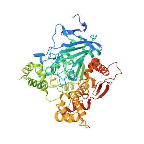
Freeze-frame inhibitor captures acetylcholinesterase in a unique conformation.

Bourne, Y.Kolb, H.C.Radic, Z.Sharpless, K.B.Taylor, P.Marchot, P.

(2004) Proc Natl Acad Sci U S A 101: 1449-1454

  • DOI: https://doi.org/10.1073/pnas.0308206100
  • Primary Citation of Related Structures:  
    1Q83, 1Q84

  • PubMed Abstract: 
  • The 1,3-dipolar cycloaddition reaction between unactivated azides and acetylenes proceeds exceedingly slowly at room temperature. However, considerable rate acceleration is observed when this reaction occurs inside the active center gorge of acetylcholinesterase (AChE) between certain azide and acetylene reactants, attached via methylene chains to specific inhibitor moieties selective for the active center and peripheral site of the enzyme ...

    The 1,3-dipolar cycloaddition reaction between unactivated azides and acetylenes proceeds exceedingly slowly at room temperature. However, considerable rate acceleration is observed when this reaction occurs inside the active center gorge of acetylcholinesterase (AChE) between certain azide and acetylene reactants, attached via methylene chains to specific inhibitor moieties selective for the active center and peripheral site of the enzyme. AChE catalyzes the formation of its own inhibitor in a highly selective fashion: only a single syn1-triazole regioisomer with defined substitution positions and linker distances is generated from a series of reagent combinations. Inhibition measurements revealed this syn1-triazole isomer to be the highest affinity reversible organic inhibitor of AChE with association rate constants near the diffusion limit. The corresponding anti1 isomer, not formed by the enzyme, proved to be a respectable but weaker inhibitor. The crystal structures of the syn1- and anti1-mouse AChE complexes at 2.45- to 2.65-A resolution reveal not only substantial binding contributions from the triazole moieties, but also that binding of the syn1 isomer induces large and unprecedented enzyme conformational changes not observed in the anti1 complex nor predicted from structures of the apoenzyme and complexes with the precursor reactants. Hence, the freeze-frame reaction offers both a strategically original approach for drug discovery and a means for kinetically controlled capture, as a high-affinity complex between the enzyme and its self-created inhibitor, of a highly reactive minor abundance conformer of a fluctuating protein template.


    Organizational Affiliation

    Ingénierie des Protéines, Centre National de la Recherche Scientifique Unité Mixte de Recherche-6560, Institut Fédératif de Recherche Jean Roche, Université de la Méditerranée, Faculté de Médecine Secteur Nord, F-13916 Marseille Cedex 20, France.


Macromolecules
Find similar proteins by:  (by identity cutoff)  |  3D Structure
Entity ID: 1
MoleculeChainsSequence LengthOrganismDetailsImage
Acetylcholinesterase
A, B
580Mus musculusMutation(s): 0 
Gene Names: Ache
EC: 3.1.1.7
UniProt & NIH Common Fund Data Resources
Find proteins for P21836 (Mus musculus)
Explore P21836 
Go to UniProtKB:  P21836
IMPC:  MGI:87876
Entity Groups  
Sequence Clusters30% Identity50% Identity70% Identity90% Identity95% Identity100% Identity
UniProt GroupP21836
Protein Feature View
Expand
  • Reference Sequence
Oligosaccharides

Help

Entity ID: 2
MoleculeChainsChain Length2D DiagramGlycosylation3D Interactions
beta-L-fucopyranose-(1-6)-2-acetamido-2-deoxy-beta-D-glucopyranose
C
2N-Glycosylation Oligosaccharides Interaction
Glycosylation Resources
GlyTouCan:  G61843VN
GlyCosmos:  G61843VN
GlyGen:  G61843VN
Small Molecules
Ligands 3 Unique
IDChainsName / Formula / InChI Key2D Diagram3D Interactions
TZ5
Query on TZ5

Download Ideal Coordinates CCD File 
F [auth A],
H [auth B]
3,8-DIAMINO-6-PHENYL-5-[6-[1-[2-[(1,2,3,4-TETRAHYDRO-9-ACRIDINYL)AMINO]ETHYL]-1H-1,2,3-TRIAZOL-5-YL]HEXYL]-PHENANTHRIDINIUM
C42 H45 N8
ISUOMOOYAOQQPZ-UHFFFAOYSA-O
 Ligand Interaction
P6G
Query on P6G

Download Ideal Coordinates CCD File 
E [auth A]HEXAETHYLENE GLYCOL
C12 H26 O7
IIRDTKBZINWQAW-UHFFFAOYSA-N
 Ligand Interaction
NAG
Query on NAG

Download Ideal Coordinates CCD File 
D [auth A],
G [auth B]
2-acetamido-2-deoxy-beta-D-glucopyranose
C8 H15 N O6
OVRNDRQMDRJTHS-FMDGEEDCSA-N
 Ligand Interaction
Binding Affinity Annotations 
IDSourceBinding Affinity
TZ5 BindingDB:  1Q83 Ki: 0.41 (nM) from 1 assay(s)
Kd: min: 4.10e-4, max: 0.03 (nM) from 2 assay(s)
PDBBind:  1Q83 Ki: 4.10e-4 (nM) from 1 assay(s)
Experimental Data & Validation

Experimental Data

  • Method: X-RAY DIFFRACTION
  • Resolution: 2.65 Å
  • R-Value Free: 0.221 
  • R-Value Work: 0.182 
  • R-Value Observed: 0.183 
  • Space Group: P 21 21 21
Unit Cell:
Length ( Å )Angle ( ˚ )
a = 79.743α = 90
b = 111.954β = 90
c = 226.577γ = 90
Software Package:
Software NamePurpose
REFMACrefinement
DENZOdata reduction
CCP4data scaling
CNSphasing

Structure Validation

View Full Validation Report


Ligand Structure Quality Assessment 


Entry History 

Deposition Data

Revision History  (Full details and data files)

  • Version 1.0: 2004-02-10
    Type: Initial release
  • Version 1.1: 2008-04-29
    Changes: Version format compliance
  • Version 1.2: 2011-07-13
    Changes: Non-polymer description, Version format compliance
  • Version 2.0: 2020-07-29
    Type: Remediation
    Reason: Carbohydrate remediation
    Changes: Advisory, Atomic model, Data collection, Derived calculations, Structure summary
  • Version 2.1: 2023-08-16
    Changes: Data collection, Database references, Refinement description, Structure summary