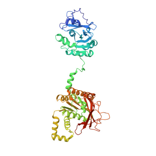
The Crystal Structure of the Bifunctional Primase-Helicase of Bacteriophage T7
Toth, E.A., Li, Y., Sawaya, M.R., Cheng, Y., Ellenberger, T.(2003) Mol Cell 12: 1113-1123
- PubMed: 14636571
- DOI: https://doi.org/10.1016/s1097-2765(03)00442-8
- Primary Citation of Related Structures:
1Q57 - PubMed Abstract:
Within minutes after infecting Escherichia coli, bacteriophage T7 synthesizes many copies of its genomic DNA. The lynchpin of the T7 replication system is a bifunctional primase-helicase that unwinds duplex DNA at the replication fork while initiating the synthesis of Okazaki fragments on the lagging strand. We have determined a 3.45 A crystal structure of the T7 primase-helicase that shows an articulated arrangement of the primase and helicase sites. The crystallized primase-helicase is a heptamer with a crown-like shape, reflecting an intimate packing of helicase domains into a ring that is topped with loosely arrayed primase domains. This heptameric isoform can accommodate double-stranded DNA in its central channel, which nicely explains its recently described DNA remodeling activity. The double-jointed structure of the primase-helicase permits a free range of motion for the primase and helicase domains that suggests how the continuous unwinding of DNA at the replication fork can be periodically coupled to Okazaki fragment synthesis.
- Department of Biological Chemistry and Molecular Pharmacology, Harvard Medical School, 240 Longwood Avenue, Boston, MA 02115, USA.
Organizational Affiliation:


