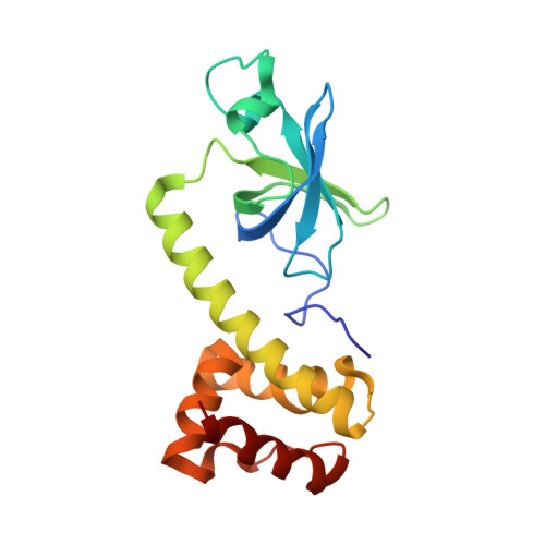
The crystal structure of the N-terminal region of the alpha subunit of translation initiation factor 2 (eIF2alpha) from Saccharomyces cerevisiae provides a view of the loop containing serine 51, the target of the eIF2alpha-specific kinases.
Dhaliwal, S., Hoffman, D.W.(2003) J Mol Biology 334: 187-195
- PubMed: 14607111 Search on PubMed
- DOI: https://doi.org/10.1016/j.jmb.2003.09.045
- Primary Citation Related Structures:
1Q46 - PubMed Abstract:
The alpha subunit of translation initiation factor 2 (eIF2alpha) is the target of specific kinases that can phosphorylate a conserved serine residue as part of a mechanism for regulating protein expression at the translational level in eukaryotes. The structure of the 20 kDa N-terminal region of eIF2alpha from Saccharomyces cerevisiae was determined by X-ray crystallography at 2.5A resolution. In most respects, the structure is similar to that of the recently solved human eIF2alpha; the rather elongated protein contains a five-stranded antiparallel beta-barrel in its N-terminal region, followed by an almost entirely helical domain. The S.cerevisiae eIF2alpha lacks a disulfide bridge that is present in the homologous protein in humans and some of the other higher eukaryotes. Interestingly, a conserved loop consisting of residues 51-65 and containing serine 51, the putative phosphorylation site, is visible in the electron density maps of the S.cerevisiae eIF2alpha; most of this functionally important loop was not observed in the crystal structure of the human protein. This loop is relatively exposed to solvent, and contains two short 3(10) helices in addition to some extended structure. Serine 51 is located at the C-terminal end of one of the 3(10) helices and near several conserved positively charged residues. The side-chain of serine 51 is sufficiently exposed so that its phosphorylation would not necessitate a substantial change in the protein structure. The structures and relative positions of residues that have been implicated in kinase binding and in the interaction with guanine nucleotide exchange factor (eIF2B) are described.
- Department of Chemistry and Biochemistry, Institute for Cellular and Molecular Biology, University of Texas at Austin, Austin, TX 78712, USA.
Organizational Affiliation: