1PXD

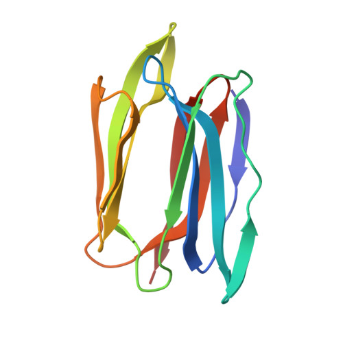
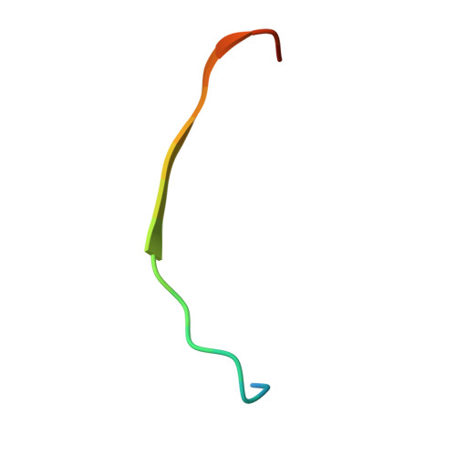
Crystal structure of the complex of jacalin with meso-tetrasulphonatophenylporphyrin.


Experimental Data Snapshot

  • Method: X-RAY DIFFRACTION
  • Resolution: 1.80 Å
  • R-Value Free: 0.233 
  • R-Value Work: 0.219 
  • R-Value Observed: 0.219 

wwPDB Validation   3D Report Full Report


Ligand Structure Quality Assessment 


This is version 1.4 of the entry. See complete history


Literature

Porphyrin binding to jacalin is facilitated by the inherent plasticity of the carbohydrate-binding site: novel mode of lectin-ligand interaction.

Goel, M.Anuradha, P.Kaur, K.J.Maiya, B.G.Swamy, M.J.Salunke, D.M.

(2004) Acta Crystallogr D Biol Crystallogr 60: 281-288

  • DOI: https://doi.org/10.1107/S0907444903026684
  • Primary Citation of Related Structures:  
    1PXD

  • PubMed Abstract: 

    The crystal structure of the complex of meso-tetrasulfonatophenylporphyrin (H(2)TPPS) with jack fruit (Artocarpus integriflora) agglutinin (jacalin) has been determined at 1.8 A resolution. A porphyrin pair is sandwiched between two symmetry-related jacalin monomers in the crystal, leading to a cross-linking network of protein molecules. Apart from the stacking interactions, H(2)TPPS also forms hydrogen bonds, some involving water bridges, with jacalin at the carbohydrate-binding site. The residues that are involved in rendering galactopyranoside specificity to jacalin undergo conformational adjustments in order to accommodate the H(2)TPPS molecule. The water molecules at the carbohydrate-binding site of jacalin cement the jacalin-porphyrin interactions, optimizing their complementarity. Interactions of porphyrin with jacalin are relatively weak compared with those observed between galactopyranoside and jacalin, perhaps because the former largely involves water-mediated hydrogen bonds. While H(2)TPPS binds to jacalin at the carbohydrate-binding site as in the case of ConA, its mode of interaction with jacalin is very different. H(2)TPPS does not enter the carbohydrate-binding cavity of jacalin. Instead, it sits over the binding site. While the porphyrin binding is mediated by replicating the hydrogen-bonding network of mannopyranoside through the sulfonate atoms in the case of ConA, the plasticity associated with the carbohydrate-binding site accommodates the pluripotent porphyrin molecule in the case of jacalin through an entirely different set of interactions.


  • Organizational Affiliation

    National Institute of Immunology, New Delhi 110067, India.


Macromolecules
Find similar proteins by:  (by identity cutoff)  |  3D Structure
Entity ID: 1
MoleculeChains Sequence LengthOrganismDetailsImage
Agglutinin alpha chain133Artocarpus integerMutation(s): 0 
UniProt
Find proteins for P18670 (Artocarpus integer)
Explore P18670 
Go to UniProtKB:  P18670
Entity Groups  
Sequence Clusters30% Identity50% Identity70% Identity90% Identity95% Identity100% Identity
UniProt GroupP18670
Sequence Annotations
Expand
  • Reference Sequence

Find similar proteins by:  Sequence   |   3D Structure  

Entity ID: 2
MoleculeChains Sequence LengthOrganismDetailsImage
Agglutinin beta-3 chain20Artocarpus integerMutation(s): 0 
UniProt
Find proteins for P18673 (Artocarpus integer)
Explore P18673 
Go to UniProtKB:  P18673
Entity Groups  
Sequence Clusters30% Identity50% Identity70% Identity90% Identity95% Identity100% Identity
UniProt GroupP18673
Sequence Annotations
Expand
  • Reference Sequence
Small Molecules
Ligands 1 Unique
IDChains Name / Formula / InChI Key2D Diagram3D Interactions
SFP
Query on SFP

Download Ideal Coordinates CCD File 
C [auth A],
D [auth A]
5,10,15,20-TETRAKIS(4-SULPFONATOPHENYL)-21H,23H-PORPHINE
C44 H34 N4 O12 S4
OVPFWIRROXFHGQ-LWQDQPMZSA-J
Binding Affinity Annotations 
IDSourceBinding Affinity
SFP Binding MOAD:  1PXD Ka: 6500 (M^-1) from 1 assay(s)
Experimental Data & Validation

Experimental Data

  • Method: X-RAY DIFFRACTION
  • Resolution: 1.80 Å
  • R-Value Free: 0.233 
  • R-Value Work: 0.219 
  • R-Value Observed: 0.219 
  • Space Group: I 2 2 2
Unit Cell:
Length ( Å )Angle ( ˚ )
a = 46.072α = 90
b = 101.89β = 90
c = 107.657γ = 90
Software Package:
Software NamePurpose
MAR345data collection
SCALEPACKdata scaling
AMoREphasing
CNSrefinement

Structure Validation

View Full Validation Report


Ligand Structure Quality Assessment 


Entry History 

Deposition Data

Revision History  (Full details and data files)

  • Version 1.0: 2004-02-03
    Type: Initial release
  • Version 1.1: 2008-04-29
    Changes: Version format compliance
  • Version 1.2: 2011-07-13
    Changes: Derived calculations, Version format compliance
  • Version 1.3: 2017-10-11
    Changes: Refinement description
  • Version 1.4: 2023-08-16
    Changes: Data collection, Database references, Derived calculations, Refinement description