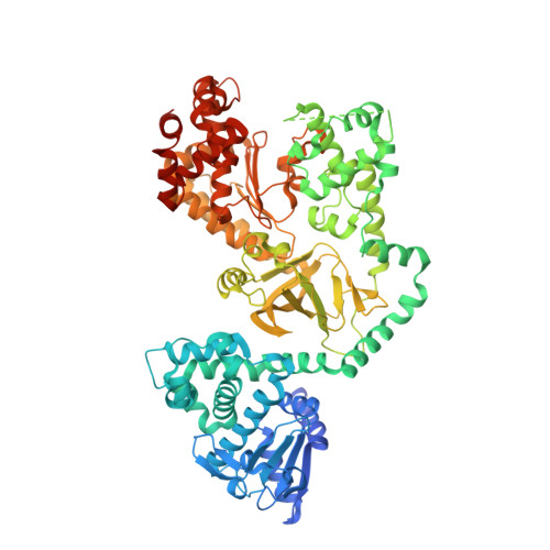
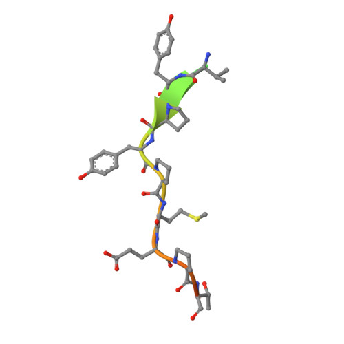
1PWV

Crystal structure of Anthrax Lethal Factor wild-type protein complexed with an optimised peptide substrate.


Experimental Data Snapshot

  • Method: X-RAY DIFFRACTION
  • Resolution: 2.85 Å
  • R-Value Free: 0.279 
  • R-Value Work: 0.231 
  • R-Value Observed: 0.231 

Starting Model: experimental
View more details

wwPDB Validation   3D Report Full Report


This is version 1.3 of the entry. See complete history


Literature

The structural basis for substrate and inhibitor selectivity of the anthrax lethal factor.

Turk, B.E.Wong, T.Y.Schwarzenbacher, R.Jarrell, E.T.Leppla, S.H.Collier, R.J.Liddington, R.C.Cantley, L.C.

(2004) Nat Struct Mol Biol 11: 60-66

  • DOI: https://doi.org/10.1038/nsmb708
  • Primary Citation of Related Structures:  
    1PWQ, 1PWU, 1PWV, 1PWW

  • PubMed Abstract: 

    Recent events have created an urgent need for new therapeutic strategies to treat anthrax. We have applied a mixture-based peptide library approach to rapidly determine the optimal peptide substrate for the anthrax lethal factor (LF), a metalloproteinase with an important role in the pathogenesis of the disease. Using this approach we have identified peptide analogs that inhibit the enzyme in vitro and that protect cultured macrophages from LF-mediated cytolysis. The crystal structures of LF bound to an optimized peptide substrate and to peptide-based inhibitors provide a rationale for the observed selectivity and may be exploited in the design of future generations of LF inhibitors.


  • Organizational Affiliation

    Division of Signal Transduction, Department of Medicine, Beth Israel Deaconess Medical Center, and Harvard Medical School, 330 Brookline Avenue, Boston, Massachusetts 02215, USA.


Macromolecules
Find similar proteins by:  (by identity cutoff)  |  3D Structure
Entity ID: 1
MoleculeChains Sequence LengthOrganismDetailsImage
Lethal factor
A, B
776Bacillus anthracisMutation(s): 0 
Gene Names: LEF OR PXO1-107
EC: 3.4.24 (PDB Primary Data), 3.4.24.83 (UniProt)
UniProt
Find proteins for P15917 (Bacillus anthracis)
Explore P15917 
Go to UniProtKB:  P15917
Entity Groups  
Sequence Clusters30% Identity50% Identity70% Identity90% Identity95% Identity100% Identity
UniProt GroupP15917
Sequence Annotations
Expand
  • Reference Sequence

Find similar proteins by:  Sequence   |   3D Structure  

Entity ID: 2
MoleculeChains Sequence LengthOrganismDetailsImage
LF20
C, D
20N/AMutation(s): 0 
Entity Groups  
Sequence Clusters30% Identity50% Identity70% Identity90% Identity95% Identity100% Identity
Sequence Annotations
Expand
  • Reference Sequence
Experimental Data & Validation

Experimental Data

  • Method: X-RAY DIFFRACTION
  • Resolution: 2.85 Å
  • R-Value Free: 0.279 
  • R-Value Work: 0.231 
  • R-Value Observed: 0.231 
  • Space Group: P 1 21 1
Unit Cell:
Length ( Å )Angle ( ˚ )
a = 96.7α = 90
b = 137.4β = 98
c = 98.3γ = 90
Software Package:
Software NamePurpose
DENZOdata reduction
SCALEPACKdata scaling
CNSrefinement
CNSphasing

Structure Validation

View Full Validation Report


Entry History 

Deposition Data

Revision History  (Full details and data files)

  • Version 1.0: 2004-02-03
    Type: Initial release
  • Version 1.1: 2008-04-29
    Changes: Version format compliance
  • Version 1.2: 2011-07-13
    Changes: Version format compliance
  • Version 1.3: 2023-08-16
    Changes: Data collection, Database references, Refinement description