


Structure of Hypothetical Protein Ywqg from Bacillus subtilis
Kim, Y., Quartey, P., Joachimiak, A.To be published.
Experimental Data Snapshot
wwPDB Validation 3D Report Full Report

Entity ID: 1 | |||||
|---|---|---|---|---|---|
| Molecule | Chains | Sequence Length | Organism | Details | Image |
| Hypothetical protein ywqG | 264 | Bacillus subtilis | Mutation(s): 7 | 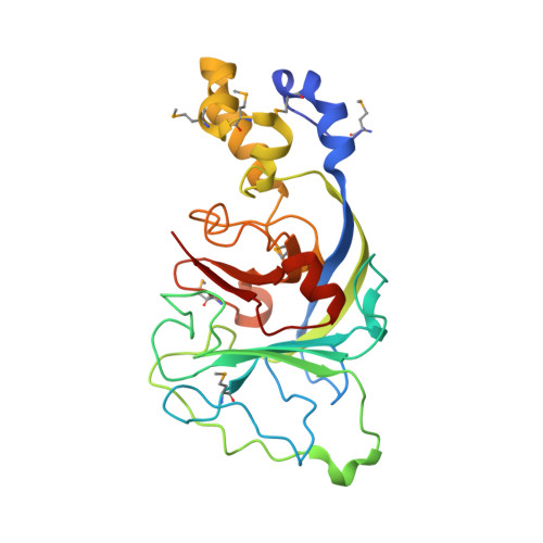 | |
UniProt | |||||
Entity Groups | |||||
| Sequence Clusters | 30% Identity50% Identity70% Identity90% Identity95% Identity100% Identity | ||||
| UniProt Group | P96719 | ||||
Sequence AnnotationsExpand | |||||
Reference Sequence | |||||
| Modified Residues 1 Unique | |||||
|---|---|---|---|---|---|
| ID | Chains | Type | Formula | 2D Diagram | Parent |
| MSE Query on MSE | A | L-PEPTIDE LINKING | C5 H11 N O2 Se |  | MET |
| Length ( Å ) | Angle ( ˚ ) |
|---|---|
| a = 38.801 | α = 90 |
| b = 88.293 | β = 103.08 |
| c = 40.103 | γ = 90 |
| Software Name | Purpose |
|---|---|
| CNS | refinement |
| SBC-Collect | data collection |
| HKL-2000 | data scaling |
| CNS | phasing |
| SOLVE | phasing |
| ARP/wARP | model building |


