1PTR

PROTEIN KINASE C DELTA CYS2 DOMAIN COMPLEXED WITH PHORBOL-13-ACETATE


Experimental Data Snapshot

  • Method: X-RAY DIFFRACTION
  • Resolution: 2.20 Å
  • R-Value Work: 0.194 
  • R-Value Observed: 0.194 

wwPDB Validation   3D Report Full Report


This is version 1.2 of the entry. See complete history


Literature

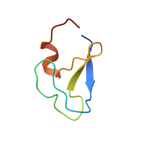
Crystal structure of the cys2 activator-binding domain of protein kinase C delta in complex with phorbol ester.

Zhang, G.Kazanietz, M.G.Blumberg, P.M.Hurley, J.H.

(1995) Cell 81: 917-924

  • DOI: https://doi.org/10.1016/0092-8674(95)90011-x
  • Primary Citation of Related Structures:  
    1PTQ, 1PTR

  • PubMed Abstract: 
  • Protein kinase Cs (PKCs) are a ubiquitous family of regulatory enzymes that associate with membranes and are activated by diacylglycerol or tumor-promoting agonists such as phorbol esters. The structure of the second activator-binding domain of PKC delta has been determined in complex with phorbol 13-acetate, which binds in a groove between two pulled-apart beta strands at the tip of the domain ...

    Protein kinase Cs (PKCs) are a ubiquitous family of regulatory enzymes that associate with membranes and are activated by diacylglycerol or tumor-promoting agonists such as phorbol esters. The structure of the second activator-binding domain of PKC delta has been determined in complex with phorbol 13-acetate, which binds in a groove between two pulled-apart beta strands at the tip of the domain. The C3, C4, and C20 phorbol oxygens form hydrogen bonds with main-chain groups whose orientation is controlled by a set of highly conserved residues. Phorbol binding caps the groove and forms a contiguous hydrophobic surface covering one-third of the domain, explaining how the activator promotes insertion of PKC into membranes.


    Organizational Affiliation

    Laboratory of Molecular Biology, National Institute of Diabetes and Digestive and Kidney Diseases, National Institutes of Health, Bethesda, Maryland 20892-0580, USA.


Macromolecules
Find similar proteins by:  (by identity cutoff)  |  3D Structure
Entity ID: 1
MoleculeChainsSequence LengthOrganismDetailsImage
PROTEIN KINASE C DELTA TYPE50Mus musculusMutation(s): 0 
Gene Names: PrkcdPkcd
EC: 2.7.1 (PDB Primary Data), 2.7.11.13 (UniProt), 2.7.10.2 (UniProt)
Membrane Entity: Yes 
UniProt
Find proteins for P28867 (Mus musculus)
Explore P28867 
Go to UniProtKB:  P28867
Entity Groups  
Sequence Clusters30% Identity50% Identity70% Identity90% Identity95% Identity100% Identity
UniProt GroupP28867
Protein Feature View
Expand
  • Reference Sequence
Small Molecules
Ligands 2 Unique
IDChainsName / Formula / InChI Key2D Diagram3D Interactions
PRB
Query on PRB

Download Ideal Coordinates CCD File 
D [auth A]13-ACETYLPHORBOL
C22 H30 O7
SDSVJYOOAPRSDA-RPCQODIISA-N
 Ligand Interaction
ZN
Query on ZN

Download Ideal Coordinates CCD File 
B [auth A],
C [auth A]
ZINC ION
Zn
PTFCDOFLOPIGGS-UHFFFAOYSA-N
 Ligand Interaction
Experimental Data & Validation

Experimental Data

  • Method: X-RAY DIFFRACTION
  • Resolution: 2.20 Å
  • R-Value Work: 0.194 
  • R-Value Observed: 0.194 
  • Space Group: C 2 2 21
Unit Cell:
Length ( Å )Angle ( ˚ )
a = 32.4α = 90
b = 63.5β = 90
c = 65.5γ = 90
Software Package:
Software NamePurpose
X-PLORmodel building
X-PLORrefinement
DENZOdata reduction
X-PLORphasing

Structure Validation

View Full Validation Report


Entry History 

Deposition Data

Revision History  (Full details and data files)

  • Version 1.0: 1995-07-31
    Type: Initial release
  • Version 1.1: 2008-03-24
    Changes: Version format compliance
  • Version 1.2: 2011-07-13
    Changes: Version format compliance