Crystal structure of human L-xylulose reductase holoenzyme: probing the role of Asn107 with site-directed mutagenesis
El-Kabbani, O., Ishikura, S., Darmanin, C., Carbone, V., Chung, R.P.-T., Usami, N., Hara, A.(2004) Proteins 55: 724-732
- PubMed: 15103634 Search on PubMed
- DOI: https://doi.org/10.1002/prot.20047
- Primary Citation Related Structures:
1PR9 - PubMed Abstract:
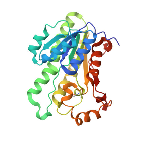
L-Xylulose reductase (XR), an enzyme in the uronate cycle of glucose metabolism, belongs to the short-chain dehydrogenase/reductase (SDR) superfamily. Among the SDR enzymes, XR shows the highest sequence identity (67%) with mouse lung carbonyl reductase (MLCR), but the two enzymes show different substrate specificities. The crystal structure of human XR in complex with reduced nicotinamide adenine dinucleotide phosphate (NADPH) was determined at 1.96 A resolution by using the molecular replacement method and the structure of MLCR as the search model. Features unique to human XR include electrostatic interactions between the N-terminal residues of subunits related by the P-axis, termed according to SDR convention, and an interaction between the hydroxy group of Ser185 and the pyrophosphate of NADPH. Furthermore, identification of the residues lining the active site of XR (Cys138, Val143, His146, Trp191, and Met200) together with a model structure of XR in complex with L-xylulose, revealed structural differences with other members of the SDR family, which may account for the distinct substrate specificity of XR. The residues comprising a recently proposed catalytic tetrad in the SDR enzymes are conserved in human XR (Asn107, Ser136, Tyr149, and Lys153). To examine the role of Asn107 in the catalytic mechanism of human XR, mutant forms (N107D and N107L) were prepared. The two mutations increased K(m) for the substrate (>26-fold) and K(d) for NADPH (95-fold), but only the N107L mutation significantly decreased k(cat) value. These results suggest that Asn107 plays a critical role in coenzyme binding rather than in the catalytic mechanism.
- Department of Medicinal Chemistry, Victorian College of Pharmacy, Monash University (Parkville Campus), Parkville, Victoria, Australia. Ossama.El-Kabbani@vcp.monash.edu.au
Organizational Affiliation: