1PK6

Globular Head of the Complement System Protein C1q


Experimental Data Snapshot

  • Method: X-RAY DIFFRACTION
  • Resolution: 1.85 Å
  • R-Value Free: 0.239 
  • R-Value Work: 0.199 
  • R-Value Observed: 0.201 

wwPDB Validation   3D Report Full Report


This is version 1.3 of the entry. See complete history


Literature

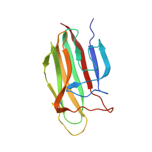
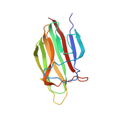
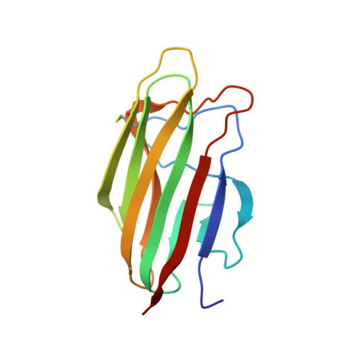
The crystal structure of the globular head of complement protein C1q provides a basis for its versatile recognition properties.

Gaboriaud, C.Juanhuix, J.Gruez, A.Lacroix, M.Darnault, C.Pignol, D.Verger, D.Fontecilla-Camps, J.C.Arlaud, G.J.

(2003) J Biol Chem 278: 46974-46982

  • DOI: https://doi.org/10.1074/jbc.M307764200
  • Primary Citation of Related Structures:  
    1PK6

  • PubMed Abstract: 
  • C1q is a versatile recognition protein that binds to an amazing variety of immune and non-immune ligands and triggers activation of the classical pathway of complement. The crystal structure of the C1q globular domain responsible for its recognition properties has now been solved and refined to 1 ...

    C1q is a versatile recognition protein that binds to an amazing variety of immune and non-immune ligands and triggers activation of the classical pathway of complement. The crystal structure of the C1q globular domain responsible for its recognition properties has now been solved and refined to 1.9 A of resolution. The structure reveals a compact, almost spherical heterotrimeric assembly held together mainly by non-polar interactions, with a Ca2+ ion bound at the top. The heterotrimeric assembly of the C1q globular domain appears to be a key factor of the versatile recognition properties of this protein. Plausible three-dimensional models of the C1q globular domain in complex with two of its physiological ligands, C-reactive protein and IgG, are proposed, highlighting two of the possible recognition modes of C1q. The C1q/human IgG1 model suggests a critical role for the hinge region of IgG and for the relative orientation of its Fab domain in C1q binding.


    Organizational Affiliation

    Laboratoire de Cristallographie et Cristallogéncse des Protéines, Institut de Biologie Structurale Jean-Pierre Ebel, Commissariat à l'Energie Atomique (CEA)-CNRS-Université Joseph Fourier, 41 rue Jules Horowitz, 38027 Grenoble Cedex 1, France.


Macromolecules
Find similar proteins by:  (by identity cutoff)  |  3D Structure
Entity ID: 1
MoleculeChainsSequence LengthOrganismDetailsImage
Complement C1q subcomponent, A chain precursorA133Homo sapiensMutation(s): 0 
Gene Names: C1QA
UniProt & NIH Common Fund Data Resources
Find proteins for P02745 (Homo sapiens)
Explore P02745 
Go to UniProtKB:  P02745
PHAROS:  P02745
Entity Groups  
Sequence Clusters30% Identity50% Identity70% Identity90% Identity95% Identity100% Identity
UniProt GroupP02745
Protein Feature View
Expand
  • Reference Sequence
Find similar proteins by:  (by identity cutoff)  |  3D Structure
Entity ID: 2
MoleculeChainsSequence LengthOrganismDetailsImage
Complement C1q subcomponent, B chain precursorB132Homo sapiensMutation(s): 0 
Gene Names: C1QB
UniProt & NIH Common Fund Data Resources
Find proteins for P02746 (Homo sapiens)
Explore P02746 
Go to UniProtKB:  P02746
PHAROS:  P02746
Entity Groups  
Sequence Clusters30% Identity50% Identity70% Identity90% Identity95% Identity100% Identity
UniProt GroupP02746
Protein Feature View
Expand
  • Reference Sequence
Find similar proteins by:  (by identity cutoff)  |  3D Structure
Entity ID: 3
MoleculeChainsSequence LengthOrganismDetailsImage
Complement C1q subcomponent, C chain precursorC129Homo sapiensMutation(s): 0 
Gene Names: C1QCC1QG
UniProt & NIH Common Fund Data Resources
Find proteins for P02747 (Homo sapiens)
Explore P02747 
Go to UniProtKB:  P02747
PHAROS:  P02747
Entity Groups  
Sequence Clusters30% Identity50% Identity70% Identity90% Identity95% Identity100% Identity
UniProt GroupP02747
Protein Feature View
Expand
  • Reference Sequence
Small Molecules
Ligands 1 Unique
IDChainsName / Formula / InChI Key2D Diagram3D Interactions
CA
Query on CA

Download Ideal Coordinates CCD File 
D [auth B]CALCIUM ION
Ca
BHPQYMZQTOCNFJ-UHFFFAOYSA-N
 Ligand Interaction
Experimental Data & Validation

Experimental Data

  • Method: X-RAY DIFFRACTION
  • Resolution: 1.85 Å
  • R-Value Free: 0.239 
  • R-Value Work: 0.199 
  • R-Value Observed: 0.201 
  • Space Group: C 1 2 1
Unit Cell:
Length ( Å )Angle ( ˚ )
a = 80.182α = 90
b = 53.222β = 112.32
c = 90.88γ = 90
Software Package:
Software NamePurpose
REFMACrefinement
XDSdata reduction
CCP4data scaling
AMoREphasing

Structure Validation

View Full Validation Report


Entry History 

Deposition Data

Revision History  (Full details and data files)

  • Version 1.0: 2003-10-21
    Type: Initial release
  • Version 1.1: 2008-04-29
    Changes: Version format compliance
  • Version 1.2: 2011-07-13
    Changes: Version format compliance
  • Version 1.3: 2018-01-31
    Changes: Advisory, Experimental preparation