1PE9 | pdb_00001pe9

MUTATIONS IN THE T1.5 LOOP OF PECTATE LYASE A


Experimental Data Snapshot

  • Method: X-RAY DIFFRACTION
  • Resolution: 1.60 Å
  • R-Value Free: 
    0.213 (Depositor), 0.213 (DCC) 
  • R-Value Work: 
    0.198 (Depositor), 0.198 (DCC) 
  • R-Value Observed: 
    0.198 (Depositor) 

Starting Model: experimental
View more details

wwPDB Validation 3D Report Full Report

Validation slider image for 1PE9

This is version 1.5 of the entry. See complete history

Literature

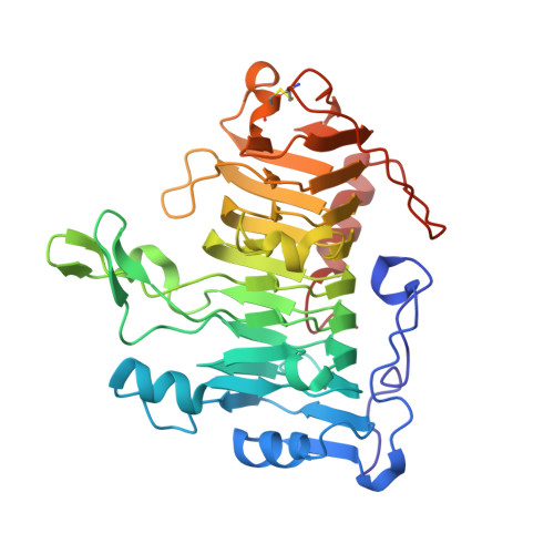
Effect of mutations in the T1.5 loop of pectate lyase A from Erwinia chrysanthemi EC16.

Dehdashti, S.J.Doan, C.N.Chao, K.L.Yoder, M.D.

(2003) Acta Crystallogr D Biol Crystallogr 59: 1339-1342

  • DOI: https://doi.org/10.1107/s0907444903011491
  • Primary Citation Related Structures: 
    1OOC, 1PE9

  • PubMed Abstract: 

    Pectate lyase A (PelA) is a pectate-degrading enzyme secreted by plant pathogens. PelA from Erwinia chrysanthemi has 61% amino-acid identity and a conserved structural similarity to pectate lyase E (PelE). Although similar in structure and sequence, the enzymatic characteristics of PelA differ from those for PelE. A structural alignment of PelA and PelE reveals differences in the T1.5 loop. The sequence of the T1.5 loop in PelA was mutated to the homologous sequence in PelE. The crystal structure of the PelA T1.5 mutant has been solved to 1.6 and 2.9 A resolution. The enzymatic and structural properties of the T1.5 mutant are discussed.


  • Organizational Affiliation
    • University of Missouri Kansas City, School of Biological Sciences, 5100 Rockhill Road, Kansas City, Missouri 64111-2499, USA.

Macromolecule Content 

  • Total Structure Weight: 77.48 kDa 
  • Atom Count: 6,230 
  • Modeled Residue Count: 722 
  • Deposited Residue Count: 722 
  • Unique protein chains: 1

Macromolecules

Find similar proteins by:|  3D Structure
Entity ID: 1
MoleculeChains  Sequence LengthOrganismDetailsImage
Pectate lyase A
A, B
361Dickeya chrysanthemiMutation(s): 4 
Gene Names: PELA
EC: 4.2.2.2
Membrane Entity: Yes 
UniProt
Find proteins for P0C1A2 (Dickeya chrysanthemi)
Explore P0C1A2 
Go to UniProtKB:  P0C1A2
Entity Groups
Sequence Clusters30% Identity50% Identity70% Identity90% Identity95% Identity100% Identity
UniProt GroupP0C1A2
Sequence Annotations
Expand
Reference Sequence

Experimental Data & Validation

Experimental Data

  • Method: X-RAY DIFFRACTION
  • Resolution: 1.60 Å
  • R-Value Free:  0.213 (Depositor), 0.213 (DCC) 
  • R-Value Work:  0.198 (Depositor), 0.198 (DCC) 
  • R-Value Observed: 0.198 (Depositor) 
Space Group: P 21 21 2
Unit Cell:
Length ( Å )Angle ( ˚ )
a = 101.797α = 90
b = 154.04β = 90
c = 51.129γ = 90
Software Package:
Software NamePurpose
CNSrefinement
DENZOdata reduction
SCALEPACKdata scaling
AMoREphasing

Structure Validation

View Full Validation Report


Entry History 

Deposition Data

Revision History  (Full details and data files)

  • Version 1.0: 2004-03-16
    Type: Initial release
  • Version 1.1: 2008-04-29
    Changes: Version format compliance
  • Version 1.2: 2011-07-13
    Changes: Version format compliance
  • Version 1.3: 2015-07-29
    Changes: Other
  • Version 1.4: 2023-08-16
    Changes: Data collection, Database references, Refinement description
  • Version 1.5: 2024-11-20
    Changes: Structure summary