


Conformational Constraint and Structural Complementarity in Thermolysin Inhibitors: Structures of Enzyme Complexes and Conclusions
Bartlett, P.A., Yusuff, N., Lindval, M.K., Holland, D., Juers, D., Matthews, B.W.To be published.
Experimental Data Snapshot
Entity ID: 1 | |||||
|---|---|---|---|---|---|
| Molecule | Chains | Sequence Length | Organism | Details | Image |
| Thermolysin | 316 | Bacillus thermoproteolyticus | Mutation(s): 0 EC: 3.4.24.27 | 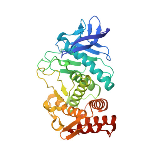 | |
UniProt | |||||
Entity Groups | |||||
| Sequence Clusters | 30% Identity50% Identity70% Identity90% Identity95% Identity100% Identity | ||||
| UniProt Group | P00800 | ||||
Sequence AnnotationsExpand | |||||
Reference Sequence | |||||
| Ligands 5 Unique | |||||
|---|---|---|---|---|---|
| ID | Chains | Name / Formula / InChI Key | 2D Diagram | 3D Interactions | |
| BR3 Download:Ideal Coordinates CCD File | G [auth A] | (6-METHYL-3,4-DIHYDRO-2H-CHROMEN-2-YL)METHYLPHOSPHINATE C11 H14 O3 P QTHZTDVLJRQOGF-JTQLQIEISA-M |  | ||
| LEU Download:Ideal Coordinates CCD File | H [auth A] | LEUCINE C6 H13 N O2 ROHFNLRQFUQHCH-YFKPBYRVSA-N |  | ||
| LEN Download:Ideal Coordinates CCD File | I [auth A] | 3-METHYLBUTAN-1-AMINE C5 H13 N BMFVGAAISNGQNM-UHFFFAOYSA-N |  | ||
| ZN Download:Ideal Coordinates CCD File | B [auth A] | ZINC ION Zn PTFCDOFLOPIGGS-UHFFFAOYSA-N |  | ||
| CA Download:Ideal Coordinates CCD File | C [auth A], D [auth A], E [auth A], F [auth A] | CALCIUM ION Ca BHPQYMZQTOCNFJ-UHFFFAOYSA-N |  | ||
| Length ( Å ) | Angle ( ˚ ) |
|---|---|
| a = 93.8 | α = 90 |
| b = 93.8 | β = 90 |
| c = 131.4 | γ = 120 |
| Software Name | Purpose |
|---|---|
| SDMS | data collection |
| SDMS | data reduction |
| TNT | refinement |
| SDMS | data scaling |
| TNT | phasing |


