1PDA | pdb_00001pda

STRUCTURE OF PORPHOBILINOGEN DEAMINASE REVEALS A FLEXIBLE MULTIDOMAIN POLYMERASE WITH A SINGLE CATALYTIC SITE


Experimental Data Snapshot

  • Method: X-RAY DIFFRACTION
  • Resolution: 1.76 Å
  • R-Value Work: 
    0.188 (Depositor), 0.172 (DCC) 
  • R-Value Observed: 
    0.188 (Depositor) 

wwPDB Validation 3D Report Full Report

Validation slider image for 1PDA

Ligand Structure Quality Assessment 


This is version 1.7 of the entry. See complete history

Literature

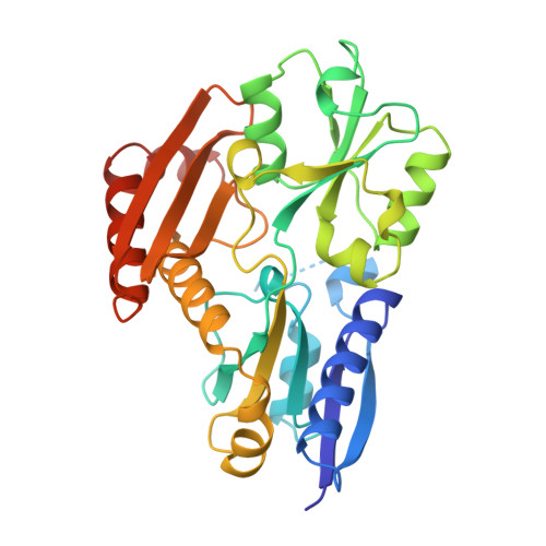
Structure of porphobilinogen deaminase reveals a flexible multidomain polymerase with a single catalytic site.

Louie, G.V.Brownlie, P.D.Lambert, R.Cooper, J.B.Blundell, T.L.Wood, S.P.Warren, M.J.Woodcock, S.C.Jordan, P.M.

(1992) Nature 359: 33-39

  • DOI: https://doi.org/10.1038/359033a0
  • Primary Citation Related Structures: 
    1PDA

  • PubMed Abstract: 

    The three-domain structure of porphobilinogen deaminase, a key enzyme in the biosynthetic pathway of tetrapyrroles, has been defined by X-ray analysis at 1.9 A resolution. Two of the domains structurally resemble the transferrins and periplasmic binding proteins. The dipyrromethane cofactor is covalently linked to domain 3 but is bound by extensive salt-bridges and hydrogen-bonds within the cleft between domains 1 and 2, at a position corresponding to the binding sites for small-molecule ligands in the analogous proteins. The X-ray structure and results from site-directed mutagenesis provide evidence for a single catalytic site. Interdomain flexibility may aid elongation of the polypyrrole product in the active-site cleft of the enzyme.


  • Organizational Affiliation
    • Department of Crystallography, Birkbeck College, University of London, UK.

Macromolecule Content 

  • Total Structure Weight: 34.37 kDa 
  • Atom Count: 2,527 
  • Modeled Residue Count: 296 
  • Deposited Residue Count: 313 
  • Unique protein chains: 1

Macromolecules

Find similar proteins by:|  3D Structure
Entity ID: 1
MoleculeChains  Sequence LengthOrganismDetailsImage
PORPHOBILINOGEN DEAMINASE313Escherichia coliMutation(s): 0 
EC: 2.5.1.61
UniProt
Find proteins for P06983 (Escherichia coli (strain K12))
Explore P06983 
Go to UniProtKB:  P06983
Entity Groups
Sequence Clusters30% Identity50% Identity70% Identity90% Identity95% Identity100% Identity
UniProt GroupP06983
Sequence Annotations
Expand
Reference Sequence

Small Molecules

Ligands 2 Unique
IDChains Name / Formula / InChI Key2D Diagram3D Interactions
DPM

Query on DPM


Download:Ideal Coordinates CCD File
B [auth A]3-[5-{[3-(2-carboxyethyl)-4-(carboxymethyl)-5-methyl-1H-pyrrol-2-yl]methyl}-4-(carboxymethyl)-1H-pyrrol-3-yl]propanoic acid
C20 H24 N2 O8
LCAXMKQKEYTFDM-UHFFFAOYSA-N
ACY

Query on ACY


Download:Ideal Coordinates CCD File
C [auth A]ACETIC ACID
C2 H4 O2
QTBSBXVTEAMEQO-UHFFFAOYSA-N

Experimental Data & Validation

Experimental Data

  • Method: X-RAY DIFFRACTION
  • Resolution: 1.76 Å
  • R-Value Work:  0.188 (Depositor), 0.172 (DCC) 
  • R-Value Observed: 0.188 (Depositor) 
Space Group: P 21 21 2
Unit Cell:
Length ( Å )Angle ( ˚ )
a = 88α = 90
b = 75.9β = 90
c = 50.5γ = 90
Software Package:
Software NamePurpose
X-PLORmodel building
PROLSQrefinement
X-PLORrefinement
X-PLORphasing

Structure Validation

View Full Validation Report


Ligand Structure Quality Assessment 


Entry History 

Deposition Data

Revision History  (Full details and data files)

  • Version 1.0: 1993-10-31
    Type: Initial release
  • Version 1.1: 2008-03-03
    Changes: Version format compliance
  • Version 1.2: 2011-07-13
    Changes: Version format compliance
  • Version 1.3: 2013-09-18
    Changes: Non-polymer description
  • Version 1.4: 2017-11-29
    Changes: Derived calculations, Other
  • Version 1.5: 2019-07-17
    Changes: Data collection, Derived calculations, Refinement description
  • Version 1.6: 2019-08-14
    Changes: Data collection, Refinement description
  • Version 1.7: 2025-03-26
    Changes: Data collection, Database references, Derived calculations, Structure summary