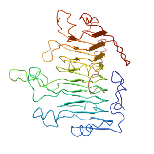
1PCL

UNUSUAL STRUCTURAL FEATURES IN THE PARALLEL BETA-HELIX IN PECTATE LYASES


Experimental Data Snapshot

  • Method: X-RAY DIFFRACTION
  • Resolution: 2.20 Å
  • R-Value Work: 0.206 
  • R-Value Observed: 0.206 

wwPDB Validation   3D Report Full Report


This is version 1.3 of the entry. See complete history


Literature

Unusual structural features in the parallel beta-helix in pectate lyases.

Yoder, M.D.Lietzke, S.E.Jurnak, F.

(1993) Structure 1: 241-251

  • DOI: https://doi.org/10.1016/0969-2126(93)90013-7
  • Primary Citation of Related Structures:  
    1PCL

  • PubMed Abstract: 

    A new type of domain structure, an all parallel beta class, has recently been observed in two pectate lyases, PelC and PelE. The atomic models have been analyzed to determine whether the new tertiary fold exhibits unusual structural features. The polypeptide backbone exhibits no new types of secondary structural elements. However, novel features occur in the amino acid side chain interactions. The side chain atoms form linear stacks that include asparagine ladders, serine stacks, aliphatic stacks, and ringed-residue stacks. A new type of beta-sandwich between parallel beta-sheets is observed with properties that are more characteristic of antiparallel beta-sheets. An analysis of the PelC and PelE structures, belonging to an all parallel beta structural class, reveals novel amino acid side chain interactions, a new type of beta-sandwich and an atypical amino acid composition of parallel beta-sheets. The findings are relevant to three-dimensional structural predictions.


  • Organizational Affiliation

    Department of Biochemistry, University of California, Riverside 92521.


Macromolecules
Find similar proteins by:  (by identity cutoff)  |  3D Structure
Entity ID: 1
MoleculeChains Sequence LengthOrganismDetailsImage
PECTATE LYASE E355Dickeya chrysanthemiMutation(s): 0 
Gene Names: PPEL748 OF PELC
EC: 4.2.2.2
UniProt
Find proteins for P04960 (Dickeya chrysanthemi)
Explore P04960 
Go to UniProtKB:  P04960
Entity Groups  
Sequence Clusters30% Identity50% Identity70% Identity90% Identity95% Identity100% Identity
UniProt GroupP04960
Sequence Annotations
Expand
  • Reference Sequence
Experimental Data & Validation

Experimental Data

  • Method: X-RAY DIFFRACTION
  • Resolution: 2.20 Å
  • R-Value Work: 0.206 
  • R-Value Observed: 0.206 
  • Space Group: P 21 21 21
Unit Cell:
Length ( Å )Angle ( ˚ )
a = 38.79α = 90
b = 91.14β = 90
c = 102.99γ = 90
Software Package:
Software NamePurpose
X-PLORmodel building
X-PLORrefinement
X-PLORphasing

Structure Validation

View Full Validation Report


Entry History 

Deposition Data

Revision History  (Full details and data files)

  • Version 1.0: 1995-02-14
    Type: Initial release
  • Version 1.1: 2008-03-24
    Changes: Version format compliance
  • Version 1.2: 2011-07-13
    Changes: Version format compliance
  • Version 1.3: 2024-02-14
    Changes: Data collection, Database references, Other