Structure of a histidine-X4-histidine zinc finger domain: insights into ADR1-UAS1 protein-DNA recognition.
Bernstein, B.E., Hoffman, R.C., Horvath, S., Herriott, J.R., Klevit, R.E.(1994) Biochemistry 33: 4460-4470
- PubMed: 8161501 Search on PubMed
- DOI: https://doi.org/10.1021/bi00181a005
- Primary Citation Related Structures:
1PAA - PubMed Abstract:
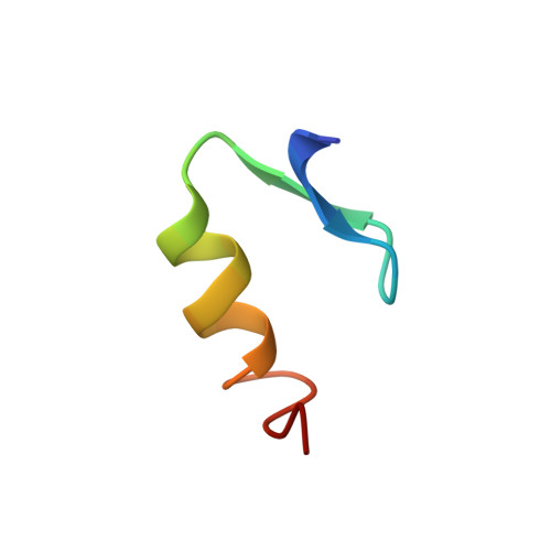
The solution structure for a mutant zinc finger peptide based on the sequence of the C-terminal ADR1 finger has been determined by two-dimensional NMR spectroscopy. The mutant peptide, called PAPA, has both proline residues from the wild-type sequence replaced with alanines. A nonessential cysteine was also replaced with alanine. The behavior of PAPA in solution implicates the prolines in the conformational heterogeneity reported earlier for the wild-type peptide [Xu, R. X., Horvath, S. J., & Klevit, R. E. (1991) Biochemistry 30, 3365-3371]. The solution structure of PAPA reveals several interesting features of the zinc finger motif. The residue immediately following the second cysteine ligand adopts a positive phi angle, which we propose is a common feature of this class of zinc fingers, regardless of whether this residue is a glycine. The NMR spectrum and resulting solution structure of PAPA suggest that a side-chain to side-chain hydrogen bond involving an arginine and an aspartic acid analogous to one observed in the Zif268 protein-DNA cocrystal structure exists in solution in the absence of DNA [Pavletich, N. P., & Pabo, C. O. (1991) Science 252, 809-817]. A model for the interaction between the two ADR1 zinc fingers and their DNA binding sites was built by superpositioning the refined solution structures of PAPA and ADR1b onto the Zif268 structure. This model offers structural explanations for a variety of mutations to the ADR1 zinc finger domains that have been shown to affect DNA-binding affinity or specificity.
- Department of Biochemistry, University of Washington, Seattle 98195.
Organizational Affiliation: