1PA1 | pdb_00001pa1

Crystal structure of the C215D mutant of protein tyrosine phosphatase 1B


Experimental Data Snapshot

  • Method: X-RAY DIFFRACTION
  • Resolution: 1.60 Å
  • R-Value Free: 
    0.204 (Depositor), 0.204 (DCC) 
  • R-Value Work: 
    0.186 (Depositor), 0.185 (DCC) 
  • R-Value Observed: 
    0.190 (Depositor) 

Starting Model: experimental
View more details

wwPDB Validation 3D Report Full Report

Validation slider image for 1PA1

This is version 1.4 of the entry. See complete history

Literature

Functional characterization and crystal structure of the C215D mutant of protein-tyrosine phosphatase-1B

Romsicki, Y.Scapin, G.Beaulieu-Audy, V.Patel, S.Becker, J.W.Kennedy, B.P.Asante-Appiah, E.

(2003) J Biological Chem 278: 29009-29015

  • DOI: https://doi.org/10.1074/jbc.M303817200
  • Primary Citation Related Structures: 
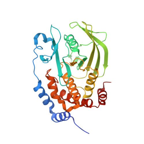
    1PA1

  • PubMed Abstract: 

    We have characterized the C215D active-site mutant of protein-tyrosine phosphatase-1B (PTP-1B) and solved the crystal structure of the catalytic domain of the apoenzyme to a resolution of 1.6 A. The mutant enzyme displayed maximal catalytic activity at pH approximately 4.5, which is significantly lower than the pH optimum of 6 for wild-type PTP-1B. Although both forms of the enzyme exhibited identical Km values for hydrolysis of p-nitrophenyl phosphate at pH 4.5 and 6, the kcat values of C215D were approximately 70- and approximately 7000-fold lower than those of wild-type PTP-1B, respectively. Arrhenius plots revealed that the mutant and wild-type enzymes displayed activation energies of 61 +/- 1 and 18 +/- 2 kJ/mol, respectively, at their pH optima. Unlike wild-type PTP-1B, C215D-mediated p-nitrophenyl phosphate hydrolysis was inactivated by 1,2-epoxy-3-(p-nitrophenoxy)propane, suggesting a direct involvement of Asp215 in catalysis. Increasing solvent microviscosity with sucrose (up to 40% (w/v)) caused a significant decrease in kcat/Km of the wild-type enzyme, but did not alter the catalytic efficiency of the mutant protein. Structurally, the apoenzyme was identical to wild-type PTP-1B, aside from the flexible WPD loop region, which was in both "open" and "closed" conformations. At physiological pH, the C215D mutant of PTP-1B should be an effective substrate-trapping mutant that can be used to identify cellular substrates of PTP-1B. In addition, because of its insensitivity to oxidation, this mutant may be used for screening fermentation broth and other natural products to identify inhibitors of PTP-1B.


  • Organizational Affiliation
    • Department of Biochemistry and Molecular Biology, Merck Frosst Centre for Therapeutic Research, Pointe-Claire, Dorval, Quebec H9R 4P8, Canada.

Macromolecule Content 

  • Total Structure Weight: 36.51 kDa 
  • Atom Count: 3,063 
  • Modeled Residue Count: 302 
  • Deposited Residue Count: 310 
  • Unique protein chains: 1

Macromolecules

Find similar proteins by:|  3D Structure
Entity ID: 1
MoleculeChains  Sequence LengthOrganismDetailsImage
Protein-tyrosine phosphatase, non-receptor type 1310Homo sapiensMutation(s): 1 
EC: 3.1.3.48
UniProt & NIH Common Fund Data Resources
Find proteins for P18031 (Homo sapiens)
Explore P18031 
Go to UniProtKB:  P18031
PHAROS:  P18031
GTEx:  ENSG00000196396 
Entity Groups
Sequence Clusters30% Identity50% Identity70% Identity90% Identity95% Identity100% Identity
UniProt GroupP18031
Sequence Annotations
Expand
Reference Sequence

Small Molecules

Ligands 2 Unique
IDChains Name / Formula / InChI Key2D Diagram3D Interactions
CL

Query on CL


Download:Ideal Coordinates CCD File
D [auth A]
E [auth A]
F [auth A]
G [auth A]
H [auth A]
D [auth A],
E [auth A],
F [auth A],
G [auth A],
H [auth A],
I [auth A]
CHLORIDE ION
Cl
VEXZGXHMUGYJMC-UHFFFAOYSA-M
MG

Query on MG


Download:Ideal Coordinates CCD File
B [auth A],
C [auth A]
MAGNESIUM ION
Mg
JLVVSXFLKOJNIY-UHFFFAOYSA-N

Experimental Data & Validation

Experimental Data

  • Method: X-RAY DIFFRACTION
  • Resolution: 1.60 Å
  • R-Value Free:  0.204 (Depositor), 0.204 (DCC) 
  • R-Value Work:  0.186 (Depositor), 0.185 (DCC) 
  • R-Value Observed: 0.190 (Depositor) 
Space Group: P 31 2 1
Unit Cell:
Length ( Å )Angle ( ˚ )
a = 88.451α = 90
b = 88.451β = 90
c = 104.356γ = 120
Software Package:
Software NamePurpose
MOSFLMdata reduction
SCALAdata scaling
CNXrefinement
CCP4data scaling
CNXphasing

Structure Validation

View Full Validation Report


Entry History 

Deposition Data

Revision History  (Full details and data files)

  • Version 1.0: 2003-08-05
    Type: Initial release
  • Version 1.1: 2008-04-29
    Changes: Version format compliance
  • Version 1.2: 2011-07-13
    Changes: Version format compliance
  • Version 1.3: 2021-10-27
    Changes: Database references, Derived calculations
  • Version 1.4: 2023-08-16
    Changes: Data collection, Refinement description