1P8T

Crystal structure of Nogo-66 Receptor


Experimental Data Snapshot

  • Method: X-RAY DIFFRACTION
  • Resolution: 3.20 Å
  • R-Value Free: 0.292 
  • R-Value Work: 0.265 
  • R-Value Observed: 0.266 

wwPDB Validation   3D Report Full Report


This is version 2.0 of the entry. See complete history


Literature

Structure and axon outgrowth inhibitor binding of the Nogo-66 receptor and related proteins

Barton, W.A.Liu, B.P.Tzvetkova, D.Jeffrey, P.D.Fournier, A.E.Sah, D.Cate, R.Strittmatter, S.M.Nikolov, D.B.

(2003) EMBO J 22: 3291-3302

  • DOI: https://doi.org/10.1093/emboj/cdg325
  • Primary Citation of Related Structures:  
    1P8T

  • PubMed Abstract: 

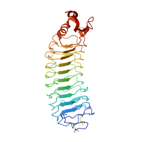
    The myelin-derived proteins Nogo, MAG and OMgp limit axonal regeneration after injury of the spinal cord and brain. These cell-surface proteins signal through multi-subunit neuronal receptors that contain a common ligand-binding glycosylphosphatidylinositol-anchored subunit termed the Nogo-66 receptor (NgR). By deletion analysis, we show that the binding of soluble fragments of Nogo, MAG and NgR to cell-surface NgR requires the entire leucine-rich repeat (LRR) region of NgR, but not other portions of the protein. Despite sharing extensive sequence similarity with NgR, two related proteins, NgR2 and NgR3, which we have identified, do not bind Nogo, MAG, OMgp or NgR. To investigate NgR specificity and multi-ligand binding, we determined the crystal structure of the biologically active ligand-binding soluble ectodomain of NgR. The molecule is banana shaped with elongation and curvature arising from eight LRRs flanked by an N-terminal cap and a small C-terminal subdomain. The NgR structure analysis, as well as a comparison of NgR surface residues not conserved in NgR2 and NgR3, identifies potential protein interaction sites important in the assembly of a functional signaling complex.


  • Organizational Affiliation

    Cellular Biochemistry and Biophysics Program, Memorial Sloan-Kettering Cancer Center, 1275 York Avenue, New York, NY 10021, USA.


Macromolecules
Find similar proteins by:  (by identity cutoff)  |  3D Structure
Entity ID: 1
MoleculeChains Sequence LengthOrganismDetailsImage
Reticulon 4 receptor285Homo sapiensMutation(s): 0 
UniProt & NIH Common Fund Data Resources
Find proteins for Q9BZR6 (Homo sapiens)
Explore Q9BZR6 
Go to UniProtKB:  Q9BZR6
PHAROS:  Q9BZR6
GTEx:  ENSG00000040608 
Entity Groups  
Sequence Clusters30% Identity50% Identity70% Identity90% Identity95% Identity100% Identity
UniProt GroupQ9BZR6
Sequence Annotations
Expand
  • Reference Sequence
Small Molecules
Ligands 2 Unique
IDChains Name / Formula / InChI Key2D Diagram3D Interactions
NDG
Query on NDG

Download Ideal Coordinates CCD File 
C [auth A]2-acetamido-2-deoxy-alpha-D-glucopyranose
C8 H15 N O6
OVRNDRQMDRJTHS-PVFLNQBWSA-N
NAG
Query on NAG

Download Ideal Coordinates CCD File 
B [auth A]2-acetamido-2-deoxy-beta-D-glucopyranose
C8 H15 N O6
OVRNDRQMDRJTHS-FMDGEEDCSA-N
Experimental Data & Validation

Experimental Data

  • Method: X-RAY DIFFRACTION
  • Resolution: 3.20 Å
  • R-Value Free: 0.292 
  • R-Value Work: 0.265 
  • R-Value Observed: 0.266 
  • Space Group: P 31 2 1
Unit Cell:
Length ( Å )Angle ( ˚ )
a = 123.958α = 90
b = 123.958β = 90
c = 120.173γ = 120
Software Package:
Software NamePurpose
DENZOdata reduction
SCALEPACKdata scaling
SHARPphasing
CNSrefinement

Structure Validation

View Full Validation Report


Entry History 

Deposition Data

Revision History  (Full details and data files)

  • Version 1.0: 2003-05-20
    Type: Initial release
  • Version 1.1: 2008-04-29
    Changes: Version format compliance
  • Version 1.2: 2011-07-13
    Changes: Non-polymer description, Version format compliance
  • Version 2.0: 2020-07-29
    Type: Remediation
    Reason: Carbohydrate remediation
    Changes: Advisory, Atomic model, Data collection, Derived calculations, Structure summary