1P4O | pdb_00001p4o

Structure of Apo unactivated IGF-1R KInase domain at 1.5A resolution.


Experimental Data Snapshot

  • Method: X-RAY DIFFRACTION
  • Resolution: 1.50 Å
  • R-Value Free: 
    0.236 (Depositor), 0.236 (DCC) 
  • R-Value Work: 
    0.209 (Depositor), 0.209 (DCC) 

wwPDB Validation 3D Report Full Report

Validation slider image for 1P4O

This is version 1.4 of the entry. See complete history

Literature

Structure of apo, unactivated insulin-like growth factor-1 receptor kinase at 1.5 A resolution.

Munshi, S.Hall, D.L.Kornienko, M.Darke, P.L.Kuo, L.C.

(2003) Acta Crystallogr D Biol Crystallogr 59: 1725-1730

  • DOI: https://doi.org/10.1107/s0907444903015415
  • Primary Citation Related Structures: 
    1P4O

  • PubMed Abstract: 

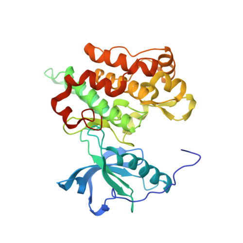
    The crystal structure of the wild-type unactivated kinase domain (IGFRK-0P) of insulin-like growth factor-1 receptor has been reported previously at 2.7 A resolution [Munshi et al. (2002), J. Biol. Chem. 277, 38797-38802]. In order to obtain a high-resolution structure, a number of variants of IGFRK-0P were prepared and screened for crystallization. A double mutant with E1067A and E1069A substitutions within the kinase-insert region resulted in crystals that diffracted to 1.5 A resolution. Overall, the structure of the mutant IGFRK-0P is similar to that of the wild-type IGFRK-0P structure, with the exception of the previously disordered kinase-insert region in the wild type having become fixed. In addition, amino-acid residues 947-952 at the N-terminus are well defined in the mutant structure. The monomeric protein structure is folded into two lobes connected by a hinge region, with the catalytic center situated at the interface of the two lobes. Two molecules of IGFRK-0P in the asymmetric unit are associated as a dimer and two different types of dimers with their ATP-binding clefts either facing towards or away from each other are observed. The current refined model consists of a dimer and 635 water molecules.


  • Organizational Affiliation
    • Department of Structural Biology, Merck Research Laboratories, West Point, PA 19486, USA. sanjeev_munshi@merck.com

Macromolecule Content 

  • Total Structure Weight: 73.31 kDa 
  • Atom Count: 5,593 
  • Modeled Residue Count: 622 
  • Deposited Residue Count: 644 
  • Unique protein chains: 1

Macromolecules

Find similar proteins by:|  3D Structure
Entity ID: 1
MoleculeChains  Sequence LengthOrganismDetailsImage
Insulin-like growth factor I receptor protein
A, B
322Homo sapiensMutation(s): 2 
Gene Names: IGF1R
EC: 2.7.1.112 (PDB Primary Data), 2.7.10.1 (UniProt)
UniProt & NIH Common Fund Data Resources
Find proteins for P08069 (Homo sapiens)
Explore P08069 
Go to UniProtKB:  P08069
PHAROS:  P08069
GTEx:  ENSG00000140443 
Entity Groups
Sequence Clusters30% Identity50% Identity70% Identity90% Identity95% Identity100% Identity
UniProt GroupP08069
Sequence Annotations
Expand
Reference Sequence

Experimental Data & Validation

Experimental Data

  • Method: X-RAY DIFFRACTION
  • Resolution: 1.50 Å
  • R-Value Free:  0.236 (Depositor), 0.236 (DCC) 
  • R-Value Work:  0.209 (Depositor), 0.209 (DCC) 
Space Group: P 1 21 1
Unit Cell:
Length ( Å )Angle ( ˚ )
a = 52.95α = 90
b = 85.56β = 99.1
c = 78.88γ = 90
Software Package:
Software NamePurpose
HKL-2000data collection
SCALEPACKdata scaling
AMoREphasing
CNSrefinement
HKL-2000data reduction

Structure Validation

View Full Validation Report


Entry History 

Deposition Data

Revision History  (Full details and data files)

  • Version 1.0: 2003-04-29
    Type: Initial release
  • Version 1.1: 2008-04-29
    Changes: Version format compliance
  • Version 1.2: 2011-07-13
    Changes: Derived calculations, Version format compliance
  • Version 1.3: 2021-10-27
    Changes: Database references
  • Version 1.4: 2024-02-14
    Changes: Data collection