Structure of porcine pancreatic phospholipase A2 at 2.6 A resolution and comparison with bovine phospholipase A2.
Dijkstra, B.W., Renetseder, R., Kalk, K.H., Hol, W.G., Drenth, J.(1983) J Mol Biology 168: 163-179
- PubMed: 6876174 Search on PubMed
- DOI: https://doi.org/10.1016/s0022-2836(83)80328-3
- Primary Citation Related Structures:
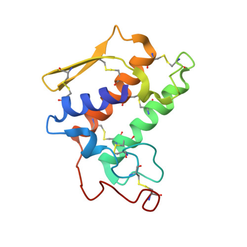
1P2P - PubMed Abstract:
The previously published three-dimensional structure of porcine pancreatic prophospholipase A2 at 3 A resolution was found to be incompatible with the structures of bovine phospholipase A2 and bovine prophospholipase A2. This was unexpected because of the very homologous amino acid sequences of these enzymes. Therefore, the crystal structure of the porcine enzyme was redetermined using molecular replacement methods with bovine phospholipase as the parent model. The structure was crystallographically refined at 2.6 A resolution by fast Fourier transform and restrained least-squares procedures to an R-factor of 0.241. The crystals appeared to contain phospholipase A2 and not prophospholipase A2. Apparently the protein is slowly converted under the crystallization conditions employed. Our investigation shows that, in contrast to the previous report, the three-dimensional structure of porcine phospholipase A2 is very similar to that of bovine phospholipase A2, including the active site. Smaller differences were observed in some residues involved in the binding of aggregated substrates. However, an appreciable conformational difference is in the loop 59 to 70, where a single substitution at position 63 (bovine Val leads to porcine Phe) causes a complete rearrangement of the peptide chain. In addition to the calcium ion in the active site, a second calcium ion is present in the crystals; this is located on a crystallographic 2-fold axis and stabilizes the interaction between two neighbouring molecules.