1OT1 | pdb_00001ot1

Bacillus circulans strain 251 Cyclodextrin glycosyl transferase mutant D135A


Experimental Data Snapshot

  • Method: X-RAY DIFFRACTION
  • Resolution: 2.00 Å
  • R-Value Free: 
    0.181 (Depositor), 0.196 (DCC) 
  • R-Value Work: 
    0.152 (Depositor) 
  • R-Value Observed: 
    0.152 (Depositor) 

Starting Model: experimental
View more details

wwPDB Validation 3D Report Full Report

Validation slider image for 1OT1

This is version 2.3 of the entry. See complete history

Literature

The fully conserved Asp residue in Conserved sequence region I of the alpha-amylase Family is crucial for the Catalytic Site Architecture and Activity

Leemhuis, H.Rozeboom, H.J.Dijkstra, B.W.Dijkhuizen, L.

(2003) FEBS Lett 541: 47-51

  • DOI: https://doi.org/10.1016/s0014-5793(03)00286-2
  • Primary Citation Related Structures: 
    1OT1, 1OT2

  • PubMed Abstract: 

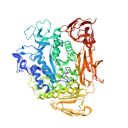
    The alpha-amylase family is a large group of starch processing enzymes [Svensson, B. (1994) Plant Mol. Biol. 25, 141-157]. It is characterized by four short sequence motifs that contain the seven fully conserved amino acid residues in this family: two catalytic carboxylic acid residues and four substrate binding residues. The seventh conserved residue (Asp135) has no direct interactions with either substrates or products, but it is hydrogen-bonded to Arg227, which does bind the substrate in the catalytic site. Using cyclodextrin glycosyltransferase as an example, this paper provides for the first time definite biochemical and structural evidence that Asp135 is required for the proper conformation of several catalytic site residues and therefore for activity.


  • Organizational Affiliation
    • Department of Microbiology, Groningen Biomolecular Sciences and Biotechnology Institute (GBB), University of Groningen, Kerklaan 30, 9751 NN Haren, The Netherlands.

Macromolecule Content 

  • Total Structure Weight: 78.3 kDa 
  • Atom Count: 6,351 
  • Modeled Residue Count: 686 
  • Deposited Residue Count: 686 
  • Unique protein chains: 1

Macromolecules

Find similar proteins by:|  3D Structure
Entity ID: 1
MoleculeChains  Sequence LengthOrganismDetailsImage
Cyclomaltodextrin glucanotransferase686Niallia circulansMutation(s): 1 
EC: 2.4.1.19
UniProt
Find proteins for P43379 (Niallia circulans)
Explore P43379 
Go to UniProtKB:  P43379
Entity Groups
Sequence Clusters30% Identity50% Identity70% Identity90% Identity95% Identity100% Identity
UniProt GroupP43379
Sequence Annotations
Expand
Reference Sequence

Oligosaccharides

Help  
Entity ID: 2
MoleculeChains Length2D Diagram GlycosylationD Interactions
alpha-D-glucopyranose-(1-4)-alpha-D-glucopyranose-(1-4)-alpha-D-glucopyranose-(1-4)-alpha-D-glucopyranose-(1-4)-alpha-D-glucopyranose-(1-4)-alpha-D-glucopyranose-(1-4)-beta-D-glucopyranose
B
7N/A
Glycosylation Resources
GlyTouCan: G99976JK
GlyCosmos: G99976JK
GlyGen: G99976JK
Entity ID: 3
MoleculeChains Length2D Diagram GlycosylationD Interactions
alpha-D-glucopyranose-(1-4)-alpha-D-glucopyranose-(1-4)-alpha-D-glucopyranose-(1-4)-beta-D-glucopyranose
C, D
4N/A
Glycosylation Resources
GlyTouCan: G69211UR
GlyCosmos: G69211UR
GlyGen: G69211UR
Entity ID: 4
MoleculeChains Length2D Diagram GlycosylationD Interactions
alpha-D-glucopyranose-(1-4)-alpha-D-glucopyranose
E
2N/A
Glycosylation Resources
GlyTouCan: G07411ON
GlyCosmos: G07411ON

Small Molecules

Ligands 4 Unique
IDChains Name / Formula / InChI Key2D Diagram3D Interactions
EPE

Query on EPE


Download:Ideal Coordinates CCD File
P [auth A]4-(2-HYDROXYETHYL)-1-PIPERAZINE ETHANESULFONIC ACID
C8 H18 N2 O4 S
JKMHFZQWWAIEOD-UHFFFAOYSA-N
MPD

Query on MPD


Download:Ideal Coordinates CCD File
I [auth A],
J [auth A],
K [auth A]
(4S)-2-METHYL-2,4-PENTANEDIOL
C6 H14 O2
SVTBMSDMJJWYQN-YFKPBYRVSA-N
ACY

Query on ACY


Download:Ideal Coordinates CCD File
L [auth A],
M [auth A],
N [auth A],
O [auth A]
ACETIC ACID
C2 H4 O2
QTBSBXVTEAMEQO-UHFFFAOYSA-N
CA

Query on CA


Download:Ideal Coordinates CCD File
F [auth A],
G [auth A],
H [auth A]
CALCIUM ION
Ca
BHPQYMZQTOCNFJ-UHFFFAOYSA-N

Biologically Interesting Molecules (External Reference) 

1 Unique

Experimental Data & Validation

Experimental Data

  • Method: X-RAY DIFFRACTION
  • Resolution: 2.00 Å
  • R-Value Free:  0.181 (Depositor), 0.196 (DCC) 
  • R-Value Work:  0.152 (Depositor) 
  • R-Value Observed: 0.152 (Depositor) 
Space Group: P 21 21 21
Unit Cell:
Length ( Å )Angle ( ˚ )
a = 116.94α = 90
b = 109.765β = 90
c = 67.75γ = 90
Software Package:
Software NamePurpose
CNSrefinement
DENZOdata reduction
SCALEPACKdata scaling
CNSphasing

Structure Validation

View Full Validation Report


Entry History 

Deposition Data

Revision History  (Full details and data files)

  • Version 1.0: 2003-06-03
    Type: Initial release
  • Version 1.1: 2008-04-29
    Changes: Version format compliance
  • Version 1.2: 2011-07-13
    Changes: Version format compliance
  • Version 2.0: 2020-07-29
    Type: Remediation
    Reason: Carbohydrate remediation
    Changes: Atomic model, Data collection, Derived calculations, Non-polymer description, Structure summary
  • Version 2.1: 2021-10-27
    Changes: Database references, Structure summary
  • Version 2.2: 2023-08-16
    Changes: Data collection, Refinement description
  • Version 2.3: 2024-11-13
    Changes: Structure summary